Clinical Measures for Preventing Contractures and Severe Scarring in Burn Wounds
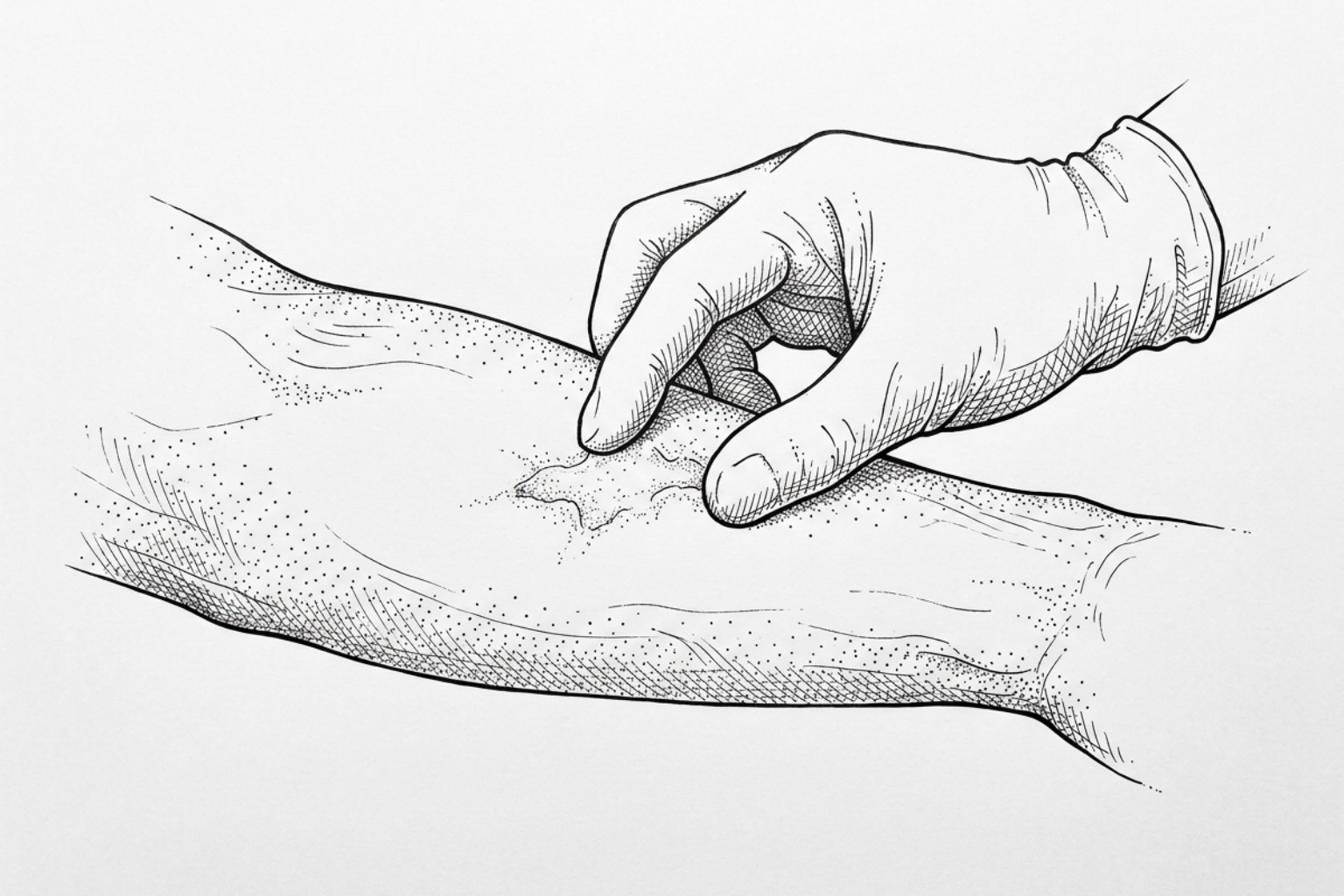
Whether a burn scars depends largely on how fast it closes. Cool the burn, keep it clean and covered, and get deep burns seen early. Once closed, silicone, compression, massage and daily SPF 30+ do the rest.
Early burn care that limits scarring
Understanding how to prevent burn scars begins in the earliest stages of injury: cool the burn with cool running water, protect it with an appropriate clean dressing, prevent infection, and seek prompt medical care for deep or extensive burns. Once the wound is fully closed, evidence suggests that silicone therapy, prescribed compression, gentle moisturization and massage, movement therapy, and daily broad-spectrum SPF 30+ protection may help scars become flatter, softer, and less noticeable.
Speed matters. Burns that heal within about 14 days are less likely to leave significant scars, while wounds that remain open for more than three weeks have a much higher risk of hypertrophic scarring and contractures. A contracture is scar tissue that tightens across a joint or body area and limits movement.
Scar prevention is not about a single cream or home remedy. It is a staged process: protect injured skin early, support orderly healing while the wound closes, then guide collagen remodeling during the months that follow. Evidence for vitamin E as a scar treatment is inconsistent, and it can irritate some people, so it should not replace clinician-guided burn care.
Clinical evidence demonstrates that staged therapeutic interventions—ranging from early wound management in second-degree burns to advanced modalities such as laser therapy for burn scars—may reduce severe scarring while protecting comfort, mobility, and long-term skin repair.
How burn scars and contractures form
The biological cascade that follows a thermal injury determines whether skin heals with minimal traces or develops severe, pathological fibrotic tissue. When thermal energy damages the dermis, it initiates a complex reparative response characterized by prolonged inflammation, dynamic cell signaling, and structural extracellular matrix remodeling. A comprehensive understanding of how scars form is necessary to appreciate why early intervention is critical.
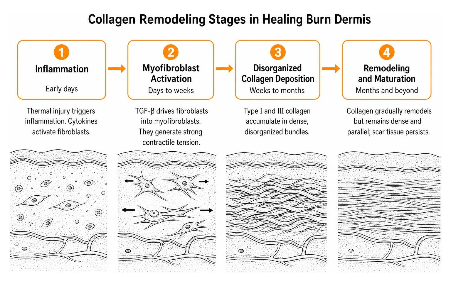
In standard cutaneous healing, inflammation resolves quickly, allowing fibroblasts to synthesize a balanced matrix of collagen. In contrast, deep burn wounds often experience sustained, dysregulated cytokine signaling. Elevated levels of transforming growth factor-beta (TGF-β) stimulate local fibroblasts, driving their persistent differentiation into contractile cells termed myofibroblasts. These myofibroblasts express alpha-smooth muscle actin, generating high mechanical tension across the wound bed.
As outlined in Comprehensive Approaches to Burn Scar Management, this persistent mechanical tension, combined with abnormal microvascular proliferation and local hypoxia, leads to the disorganized accumulation of type I and type III collagen fibers. Instead of arranging in the pliable, basket-weave lattice characteristic of uninjured dermis, the newly formed collagen bundles organize into dense, parallel nodular arrays. This excessive matrix deposition produces rigid, unyielding scar tissue.
Types of burn scars
Cutaneous scarring after thermal injury falls into distinct pathological categories. Understanding the specific scar types helps clinicians select appropriate prophylactic and therapeutic interventions:
- Hypertrophic Scars: These are raised, rigid, erythematous (red or purple) scars that remain strictly confined within the boundaries of the original burn margin. They typically develop within weeks to months following thermal trauma and are characterized by high cellularity, dense collagen nodules, and persistent microvascular hyperperfusion.
- Keloid Scars: While sharing certain histological features with hypertrophic scars, keloids represent an autonomous fibroproliferative disorder. They extend beyond the original anatomic margins of the thermal injury, invading adjacent healthy tissue. Keloids rarely regress spontaneously and show higher recurrence rates following surgical excision.
- Burn Scar Contractures: Contractures develop when excessive myofibroblast contraction and dense extracellular matrix deposition occur over or adjacent to articulating joints, flexural creases, or facial structures. The resulting structural shortening of the cutaneous envelope restricts passive and active range of motion, creating substantial functional impairment.
Clinicians evaluate these tissue changes using standardized assessment tools, notably the Vancouver Scar Scale (VSS) and the Patient and Observer Scar Assessment Scale (POSAS). These clinical metrics quantify vascularity, pigmentation, pliability, and scar height to guide therapeutic adjustments throughout rehabilitation.
The 14-Day Epithelialization Window and Healing Timelines
Clinical outcomes demonstrate a direct correlation between the duration required for complete re-epithelialization and the ultimate incidence of severe scarring. Current clinical consensus establishes a 14-day threshold as a primary prognostic marker:
- Under 14 Days: Burns that achieve complete epithelialization within 14 days (typically superficial and superficial partial-thickness burns) generally exhibit minimal risk of long-term hypertrophic scarring or contracture development.
- 14 to 21 Days: Wounds closing within this intermediate window carry an increased risk of textural irregularity and mild to moderate hypertrophic scarring, warranting close surveillance.
- Greater than 21 Days: Wounds that take longer than three weeks to heal (primarily deep partial-thickness and full-thickness injuries) carry an exceptionally high risk of pathological hypertrophic scar formation, often exceeding 70% to 80% incidence without aggressive intervention (Walker & King, 2023).
When thermal injuries extend deep into the reticular dermis, destruction of dermal appendages (such as hair follicles and sweat glands) impairs spontaneous re-epithelialization. The resulting extended inflammatory phase amplifies fibroblastic hyperactivity, reinforcing the clinical objective: accelerate safe wound closure to minimize the inflammatory duration.
First aid and early wound care
Immediate, evidence-based acute care establishes the foundation for scar prevention. The initial minutes and hours following thermal trauma dictate the survival of marginal tissue within the wound bed.
According to guidelines from major clinical centers, such as those detailed in Burns - Diagnosis and treatment - Mayo Clinic, immediate first aid requires cooling the burned area under cool running tap water (approximately 12°C to 18°C) for 10 to 20 minutes. Ice or freezing water must never be applied, as extreme cold induces severe vasoconstriction and secondary tissue necrosis, compounding dermal damage.
Jackson's burn model illustrates that thermal wounds comprise three distinct concentric zones:
- Zone of Coagulation: The central area of maximal thermal absorption characterized by irreversible cellular necrosis and protein denaturation.
- Zone of Stasis: The intermediate, vulnerable zone of compromised microvascular perfusion and marginal tissue viability. Without rapid cooling, adequate systemic hydration, and infection control, this zone easily converts to non-viable necrotic tissue, converting a superficial partial-thickness burn into a deep partial- or full-thickness injury.
- Zone of Hyperemia: The outermost peripheral area exhibiting increased perfusion and inflammation, which typically recovers fully under standard care.
In extensive thermal injuries, systemic fluid resuscitation using established clinical protocols (such as the Parkland or Modified Brooke formulas) maintains organ and microvascular perfusion. Adequate perfusion preserves the zone of stasis, shortens healing time, and reduces long-term scarring risks.
Moist dressings for burns
Maintaining a physiologic, moist wound environment is essential for rapid re-epithelialization. When a wound bed is exposed to open air and allowed to dry, keratinocytes must migrate beneath a hard, non-viable crust (eschar or scab) to re-establish tissue integrity. This deep migration slows epithelialization and intensifies inflammatory signaling.
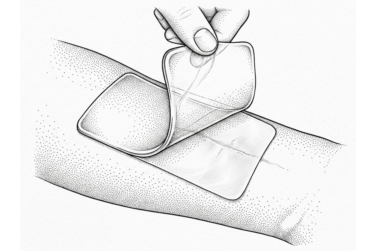
A moist healing environment provides optimal conditions for epidermal cell migration across the viable wound surface. Implementing appropriate clinical strategies to protect healing skin involves selecting advanced wound dressings tailored to the exudate level and depth of the burn:
- Semi-Occlusive and Hydrogel Dressings: For superficial to partial-thickness burns, hydrogels and polyurethane films maintain optimal hydration, cool the underlying sensory nerve endings to ease pain, and support autolytic debridement.
- Petroleum-Based Ointments: Sterile petroleum jelly provides an inert, non-sensitizing barrier that prevents trans-epidermal water loss, accelerates cell migration, and prevents secondary trauma during dressing changes.
- Non-Adherent Silicone Dressings: Porous silicone contact layers protect delicate, newly generated epithelial sheets from mechanical shear forces during daily dressing changes.
Dressings must be changed cleanly and routinely, inspecting the wound bed daily to maintain moisture balance without causing maceration of surrounding healthy tissue.
Cleaning the wound and preventing infection
Infection is a major cause of prolonged inflammation and delayed epithelialization, significantly increasing the risk of hypertrophic scarring. Bacterial overgrowth (particularly by Pseudomonas aeruginosa or Staphylococcus aureus) releases destructive matrix metalloproteinases and toxins that break down regenerating tissue.
Clinical wound management requires meticulous, gentle cleansing using mild soaps or isotonic saline solutions. Non-viable, loose necrotic tissue must be debrided carefully by clinical professionals to eliminate bacterial growth substrates. For deep partial-thickness burns with elevated microbial risk, topical antimicrobial agents—including silver sulfadiazine, silver-impregnated dressings, or mafenide acetate—may be utilized selectively to suppress microbial proliferation while protecting viable cellular elements.
Pressure, massage and topical treatments
Once complete re-epithelialization occurs and the fragile epidermal barrier is re-established, proactive scar prevention shifts toward mechanical and topical therapies. A structured approach to burn scar treatment highlights the multimodal nature of modern rehabilitation.
| Intervention Modality | Mechanism of Action | Recommended Protocol | Primary Clinical Indication |
|---|---|---|---|
| Medical-Grade Silicone | Stratum corneum hydration, reduced capillary activity, decreased collagen synthesis | 12 to 24 hours daily for 3 to 6+ months | Immature, erythematous scars; early prophylactic care |
| Compression Therapy | Capillary occlusion, reduced fibroblast proliferation, parallel collagen realignment | 15–30 mmHg gradient; 23 hours/day for 6–18 months | Deep partial- to full-thickness burns healing >14–21 days |
| Scar Massage & Stretching | Mechanotransduction, tissue pliability enhancement, relief of joint restriction | 2–3 sessions daily, 5–10 minutes per site | Contracture prevention, joint mobility, pruritus reduction |
Compression and silicone
Compression therapy and topical silicone represent standard physical modalities in burn scar rehabilitation.
Continuous external pressure applied via custom-fitted garments (typically calibrated to maintain 15 to 30 mmHg of gradient compression) works by reducing capillary flow to active scar tissue. This controlled local ischemia downregulates fibroblast metabolism, reduces local TGF-β concentrations, and encourages newly synthesized collagen fibers to realign parallel to the epidermal surface rather than forming dense nodules. For maximum therapeutic benefit, compression garments are worn approximately 23 hours per day—removed solely for hygiene—for a duration of 6 to 18 months, or until the scar demonstrates complete vascular and cellular maturation.
Concurrently, topical silicone gel and medical-grade silicone gel sheets deliver targeted epidermal occlusion. In clinical evaluations of hypertrophic scar silicone gel applications, silicone's primary therapeutic mechanism is the restoration of the stratum corneum barrier. By minimizing trans-epidermal water loss (TEWL), silicone normalizes signaling between surface keratinocytes and underlying dermal fibroblasts, dampening excessive matrix synthesis. Silicone therapy should be applied consistently for 12 to 24 hours daily for at least 8 to 12 weeks.
Massage and preventing contractures
Mechanical manipulation of newly healed burn tissue prevents cutaneous adhesions to underlying fascia and preserves soft tissue compliance.
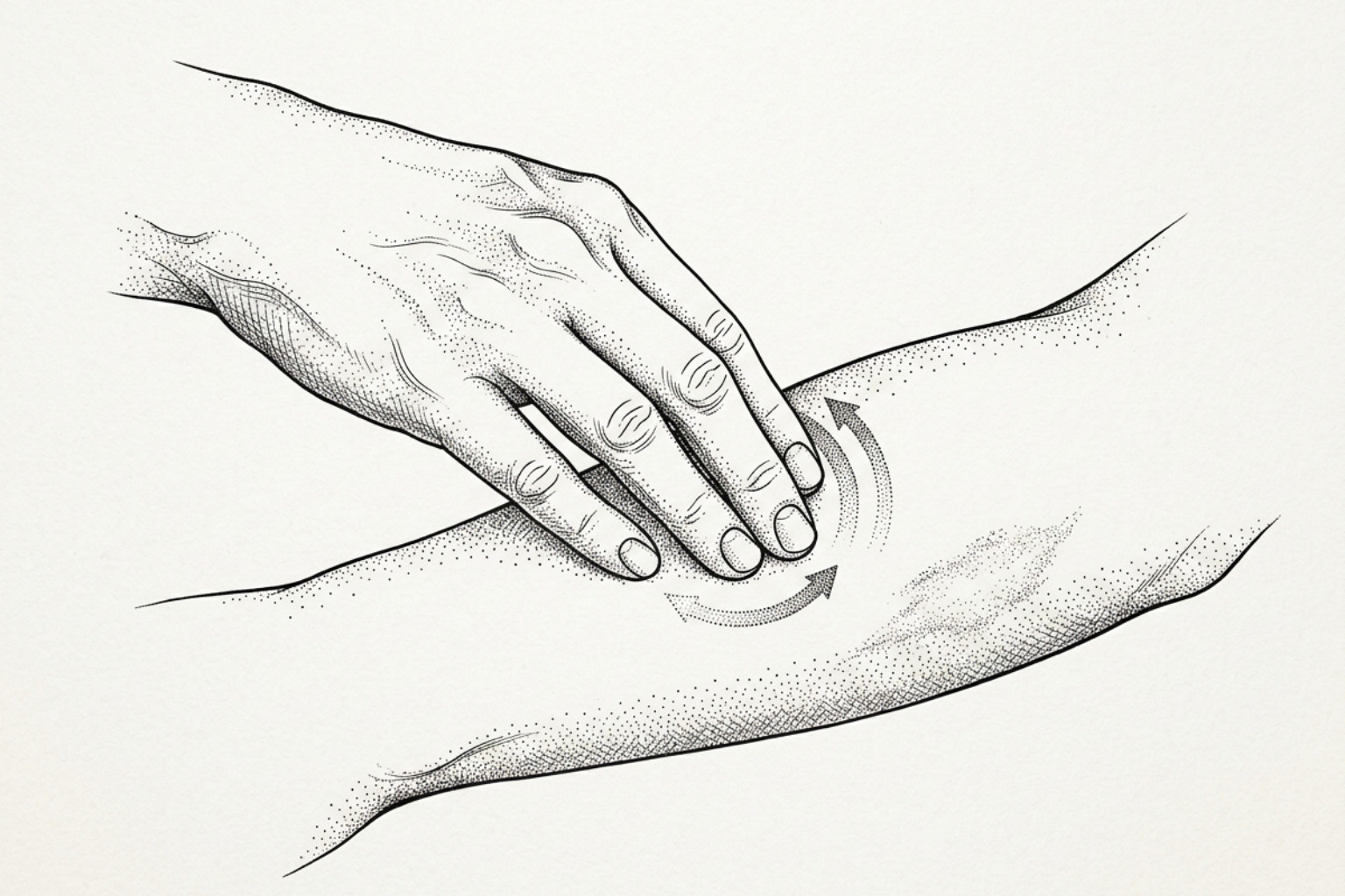
Once the neo-epidermis is sufficiently durable to tolerate shear forces without blister formation, targeted massage should be introduced. As detailed in clinical guidelines for scar tissue massage, applying gentle, circular, perpendicular, and skin-rolling friction forces for 5 to 10 minutes, two to three times daily, helps break up immature, rigid cross-linked collagen bundles.
For thermal injuries traversing or adjacent to articulating joints, scar massage must be integrated with active and passive range-of-motion (ROM) physical therapy. Consistent stretching exercises, performed 5 to 6 times daily, counteract ongoing myofibroblast-mediated contractures. When necessary, custom-molded static or dynamic splints are worn overnight or between exercise sessions to hold healing tissues at maximal functional elongation.
Sun protection and preventing dark marks
Newly regenerated skin over a healed burn injury is susceptible to post-inflammatory hyperpigmentation (PIH) and persistent erythema. The fragile, immature epidermal barrier contains unstable melanocytes that react strongly to ultraviolet radiation (UVR), which can lead to long-term, mottled discoloration.
According to the dermatology consensus presented in How To Prevent Scarring: Dos and Don’ts, strict photoprotection is mandatory for a minimum of 6 to 12 months post-closure. Survivors must apply broad-spectrum sunscreen with an SPF of 30 or higher, reapplying every two hours during outdoor exposure. Physical blocking agents containing zinc oxide or titanium dioxide are particularly suitable, as they reflect UV radiation without irritating delicate, healing skin. In addition, physical barriers such as tightly woven, UV-protective garments should be used whenever feasible.
Procedures, nutrition and scar maturation
Burn scar remodeling is an extended biological process lasting between 12 and 18 months, during which the initial, highly vascular tissue gradually matures into a stable cutaneous scar. Optimal outcomes often require specialized clinical interventions and metabolic support.
Early laser and procedures
Advanced energy-based devices play a significant role in modern burn scar management. Rather than delaying procedural intervention until full maturity, evidence supports initiating targeted laser treatments within 1 to 3 months of wound closure:
- Pulsed Dye Laser (PDL - 585 to 595 nm): By targeting the hemoglobin absorption peak within hypervascular scar tissue, PDL photocoagulates abnormal microvasculature. This reduces visible erythema, lowers local metabolic activity, relieves severe pruritus, and limits further hypertrophic growth.
- Fractional Ablative CO2 Lasers (10,600 nm): As examined in clinical overviews of burn scar laser treatment, fractional CO2 lasers create microscopic vertical thermal treatment zones that penetrate deep into the reticular dermis. This thermal injury stimulates controlled neocollagenesis and matrix remodeling, improving tissue pliability, releasing superficial tension, and reducing scar thickness.
- Intralesional Corticosteroids: For recalcitrant, highly raised hypertrophic bands, precise intralesional injections of triamcinolone acetonide (often combined with 5-fluorouracil) can be administered to suppress fibroblast proliferation and accelerate matrix degradation.
Systemic Nutrition and Metabolic Support
Severe thermal trauma induces a profound hypermetabolic state that can persist for months, accelerating protein catabolism and increasing caloric demands. Adequate systemic nutrition is necessary to support tissue repair and orderly collagen synthesis.
Maintaining optimal healing requires a balanced, nutrient-dense diet:
- High Dietary Protein: Protein provides essential amino acids (such as proline, glycine, and arginine) required for cellular repair and structural collagen remodeling, reinforcing the importance of adequate protein in wound healing.
- Ascorbic Acid (Vitamin C): Serves as an obligate cofactor in the enzymatic hydroxylation of lysine and proline residues, a biochemical step required for stable collagen cross-linking and tensile strength.
- Zinc and Micronutrients: Zinc functions as a critical structural component for numerous enzymes and transcription factors involved in re-epithelialization and immune defense, alongside other essential vitamins and micronutrients supporting tissue regeneration.
Common questions about preventing burn scars
How long do burn scars take to fully mature?
Burn scars typically require 12 to 18 months—and in some deep thermal injuries, up to two to three years—to achieve complete physiological maturation. During this timeframe, the scar progresses from an active, vascular, hypercellular state (peaking in thickness, firmness, and redness around 6 months) toward a stable equilibrium characterized by diminished blood flow, reduced cellularity, improved pliability, and a softer texture closer to the surrounding skin tone.
When should compression garments and silicone sheets be initiated?
Compression therapy and silicone gel applications should begin as soon as the wound bed achieves complete re-epithelialization—meaning the epidermal surface is intact, durable, free of open blisters or raw drainage, and cleared by the managing burn specialist or clinician. Initiating these therapies early during the immature scar phase helps control fibroblastic activity and prevents excessive extracellular matrix deposition before collagen nodules become firmly established.
Can natural remedies like aloe vera or vitamin E prevent severe burn scarring?
While pure, medical-grade aloe vera extract provides soothing hydration for minor, superficial first-degree burns, there is no high-quality clinical evidence indicating that it prevents hypertrophic scarring or contractures in deeper thermal injuries. Topical vitamin E (tocopherol) has shown inconsistent, conflicting results in clinical studies; in addition, research indicates it can cause contact dermatitis and localized irritation in up to 20% to 30% of users. Evidence-based care centers on clinical modalities: moist wound healing, medical-grade silicone, compression garments, UV photoprotection, and movement therapy.
Key takeaways
Minimizing long-term complications and severe scarring after a burn injury requires a disciplined, multimodal approach. The window for intervention begins immediately at the time of thermal trauma: cooling the burn safely, preserving marginal tissue within the zone of stasis, maintaining a moist healing environment, and closing the wound rapidly are the first lines of defense.
Once the epidermal surface is restored, consistent application of medical-grade silicone, custom compression garments, daily targeted scar massage, active joint mobilization, and strict photoprotection guide the remodeling phase. For deep or extensive burns, early consultation with specialized burn rehabilitation teams ensures access to advanced modalities such as fractional laser therapy, dynamic splinting, or surgical revision. For more detailed clinical information on specific burn depths and long-term functional recovery, consult clinical overviews on burn scar types and rehabilitation.
This content is for informational purposes only and does not constitute medical advice. Consult a qualified healthcare professional for diagnosis and treatment.
Works Cited
- Walker NJ, King KC. “Acute and Chronic Thermal Burn Evaluation and Management.” StatPearls, 2023.
- Nedelec B, Carter A, Forbes L, et al. “Practice Guidelines for the Application of Nonsilicone or Silicone Gels and Gel Sheets After Burn Injury.” Journal of Burn Care & Research, 2015.
- Monstrey S, Middelkoop E, Vranckx JJ, et al. “Updated scar management practical guidelines: non-invasive and invasive measures.” Journal of Plastic, Reconstructive & Aesthetic Surgery, 2014.
- Orgill DP, Ogawa R. “Current methods of burn reconstruction.” Plastic and Reconstructive Surgery, 2013.