How to Manage Second-Degree Burns to Minimize the Risk of Long-Term Scars
Whether a second-degree burn leaves a scar depends mostly on how fast it closes. Wounds healed within 14 days scar under 10% of the time; past 21 days the risk climbs sharply. Here is what changes that.
Understanding Burn Depth and Pathophysiology
To minimize the risk of second-degree burn scars, immediate and post-closure clinical management must focus on halting tissue damage and promoting rapid re-epithelialization. Evidence-based steps include:
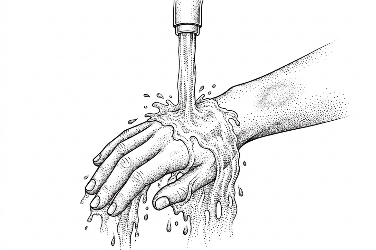
- Prompt Cooling: Cool the burn with running tap water (12°C to 25°C) for at least 20 minutes within 3 hours of injury.
- Blister Preservation: Leave intact blister roofs intact to serve as sterile biological barriers against infection.
- Moist Wound Healing: Use advanced dressings (such as hydrocolloids or silicone foams) to achieve complete wound closure within 14 to 21 days.
- Post-Closure Care: Apply medical-grade silicone sheeting, broad-spectrum sun protection, and pressure therapy once re-epithelialization occurs.
When thermal energy damages cutaneous tissue, the microenvironment undergoes complex pathological transformations. Second-degree injuries, clinically termed partial-thickness burns, extend beyond the superficial epidermis into the underlying dermis. The survival of skin appendages within this dermal reservoir—specifically hair follicles, sebaceous glands, and sweat ducts—determines the capacity for spontaneous re-epithelialization.
In burn pathophysiology, Jackson's burn wound model describes three concentric zones of tissue injury:
- Zone of Coagulation: The central area of irreversible tissue necrosis where cellular proteins have denatured.
- Zone of Stasis: The surrounding tissue characterized by compromised microvascular perfusion. This zone is vulnerable to progressive ischemia, desiccation, edema, and secondary necrosis if not properly resuscitated.
- Zone of Hyperemia: The outermost perimeter exhibiting increased blood flow and robust inflammatory response, typically resolving without tissue loss.

Preserving the viability of the zone of stasis is the primary clinical objective during acute management. If microvascular thrombosis occurs, superficial injuries can convert into deeper dermal wounds, drastically increasing pathological collagen deposition. Understanding how scars form requires examining this biological shift: prolonged inflammation signals dermal fibroblasts to synthesize disorganized extracellular matrix (ECM) components rather than normal, pliable basket-weave collagen bundles (Ji et al., 2024).
Superficial vs. Deep Partial-Thickness Wounds
Second-degree burns represent a spectrum rather than a uniform injury depth. Clinical guidelines distinguish between superficial partial-thickness, shallow deep, and profound deep dermal injuries based on anatomical depth, capillary refill, and sensory preservation.
According to clinical resources like the Cleveland Clinic 2nd-Degree Burn Overview, superficial partial-thickness burns damage the epidermis and the upper layer of the dermis (the papillary dermis). These wounds typically present as moist, bright red or pink surfaces with intact or ruptured thin-walled blisters. Because sensory nerve endings remain intact, these wounds are exquisitely painful. Capillary refill remains brisk upon gentle pressure, reflecting preserved microvascular integrity.
In contrast, deep partial-thickness burns involve the deeper reticular dermis. These wounds often appear mottled pink, white, or waxy and dry, with thick-walled blisters. Sensation is frequently reduced due to the destruction of superficial nerve networks, and capillary refill is sluggish or absent. Distinguishing between shallow deep and profound deep wounds remains clinically challenging: international data indicates that even experienced burn specialists achieve only a 50% to 70% diagnostic accuracy rate for predicting 3-week wound healing based on visual assessment alone (Ji et al., 2024).
Why Healing Time Predicts Second-Degree Burn Scars
The duration required for a partial-thickness wound to achieve complete re-epithelialization serves as the most dependable clinical predictor for the development of pathological raised or hypertrophic tissue. Prolonged open wounds sustain chronic cellular signaling pathways involving transforming growth factor-beta (TGF-β), which recruits active myofibroblasts and promotes excessive deposition of dense type I collagen fibers.
| Burn Depth Classification | Dermal Layer Affected | Average Re-epithelialization Time | Hypertrophic Scar Risk |
|---|---|---|---|
| Superficial Partial-Thickness | Epidermis and Papillary Dermis | 7 to 14 days | < 10% (minimal risk; pigment shifts possible) |
| Shallow Deep Partial-Thickness | Extends into Mid-Reticular Dermis | 14 to 21 days | Approximately 30% |
| Profound Deep Partial-Thickness | Deep Reticular Dermis | > 21 days (often 4 to 6 weeks) | 70% to 80% |
Clinical observations demonstrate that crossing the 21-day threshold shifts the wound healing trajectory from physiological tissue repair toward fibroproliferative remodeling. When epithelial closure requires more than 3 weeks, aggressive scar management strategies become necessary to mitigate contracture formation and functional impairment.
Immediate Care and Wound Interventions to Prevent Tissue Deepening
The primary goal of prehospital and acute clinical burn management is preventing secondary tissue necrosis within the zone of stasis. Second-degree burns account for 85.4% of all burn cases globally, with 56.3% involving less than 10% of the total body surface area (TBSA) (Ji et al., 2024). Rapid, appropriate first aid plays a decisive role in halting thermal injury progression.
Prehospital Cooling and Blister Preservation
The 2024 Consensus on the Treatment of Second-Degree Burn Wounds outlines standard protocols for prehospital first aid (Ji et al., 2024):
- Thermal Dissipation: Initiate cooling with running tap water (ideal temperature between 12°C and 25°C) as soon as possible, ideally within 3 hours of injury.
- Duration: Maintain active cooling for at least 20 minutes, or until acute pain subsides. Studies indicate this intervention halts thermal transfer, decreases local edema, and significantly reduces the need for subsequent skin grafting (Ji et al., 2024).
- Precautions: Never apply ice, iced water, or freezing compresses directly to burn wounds. Extreme cold causes severe reactive vasoconstriction, worsening microvascular ischemia and causing cold-induced tissue injury. In pediatric patients or those with large burn areas, monitor core body temperature closely to avoid systemic hypothermia.
- Blister Roof Preservation: Keep intact blister roofs intact during prehospital care. The blister roof acts as a sterile, non-adherent biological dressing that protects delicate basal keratinocytes, reduces neuropathic pain, and decreases infection risk. If large blisters impair joint articulation, fluid can be sterilely aspirated by a medical provider while leaving the overlying epidermis in place.
Advanced Dressings, Enzymatic Debridement, and Growth Factors
Managing the acute wound bed requires maintaining a balanced moisture level, controlling bacterial colonization, and accelerating cellular proliferation. For deep partial-thickness wounds, advanced topical modalities help minimize prolonged inflammatory responses:
- Advanced Dressings: Hydrocolloid dressings, silicone-coated polyurethane foams, and silver-impregnated barrier dressings sustain a moist wound environment that facilitates keratinocyte migration while managing wound exudate.
- Enzymatic Debridement: Applying targeted enzymatic agents, such as collagenase or bromelain-based formulations, allows for the selective digestion of devitalized necrotic tissue without destroying viable dermal elements. Research indicates enzymatic debridement may promote cleaner wound beds faster than traditional conservative ointment therapy (Zanganeh et al., 2022).
- Recombinant Growth Factors: Emerging clinical data supports using topical growth factors to stimulate tissue regeneration. A randomized controlled trial evaluating pediatric second-degree burns demonstrated that basic fibroblast growth factor (bFGF) significantly shortened the time to cessation of wound exudation (13.8 ± 2.4 days versus 17.5 ± 3.1 days in controls) and eliminated hypertrophic scar formation at 1-year follow-up (Ji et al., 2024). Similarly, recombinant human epidermal growth factor (rhEGF) has been shown to enhance cellular proliferation and optimize the collagen type I/III ratio during dermal tissue expansion (He et al., 2025).
Clinical Protocols to Manage and Treat Second-Degree Burn Scars
Once complete re-epithelialization occurs, the wound enters the remodeling phase, which can last from 12 to 24 months. During this period, the balance between collagen synthesis and degradation dictates scar texture, elevation, and pliability. Comprehensive non-operative strategies outlined in clinical resources such as the non-surgical burn scars complete guide and best burn scars treatment guide provide early lines of defense against hypertrophic remodeling.
Advanced Laser and Topical Therapies for Second-Degree Burn Scars
Clinical protocols for managing mature or developing second-degree burn scars combine physical compression, hydration barrier restoration, and photothermal remodeling:
- Silicone Gel Sheeting: Medical-grade silicone sheets or gels are the established first-line non-invasive standard of care. Silicone normalizes stratum corneum hydration, reducing transepidermal water loss (TEWL) and signaling down-regulation of cytokine production by fibroblasts.
- Pressure Garment Therapy (PGT): Providing continuous gradient pressure (typically 15–25 mmHg) for 23 hours per day compresses local capillary beds, limits fibroblast metabolism, and encourages parallel alignment of newly formed collagen bundles.
- Vascular Laser Modalities (Pulsed Dye Laser - 595 nm): As detailed in the burn scar laser treatment guide, pulsed dye laser (PDL) targets the microvascular hemoglobin within erythematous scars. Photocoagulating abnormal vessels reduces scar hyperemia, curbs nutrient delivery to active myofibroblasts, and relieves persistent pruritus.
- Fractional Ablative Lasers (CO2 / Erbium:YAG): Microscopic treatment zones vaporize narrow columns of dense, rigid scar tissue. This controlled thermal micro-injury stimulates normal collagen remodeling, releases physical tension across tight bands, and improves tissue pliability (He et al., 2025).
Indications for Surgical Debridement and Skin Grafting
When deep partial-thickness burns fail to show signs of spontaneous healing by 14 to 21 days, surgical options are evaluated to reduce prolonged inflammation and prevent extensive scarring:
- Tangential Excision and Split-Thickness Skin Grafting (STSG): In profound deep burns involving complex cosmetic or functional units (such as the hands, neck, or flexor joints), early tangential excision of necrotic dermis followed by STSG placement reduces healing time, limits hypertrophic scarring, and preserves range of motion.
- Conservative Trunk Management: For deep partial-thickness burns on the back, trunk, or buttocks, conservative nonsurgical dressing regimens may occasionally produce lower Vancouver Scar Scale (VSS) scores than skin grafting, particularly when donor skin sites are limited (Zanganeh et al., 2022). Constant natural pressure from body weight when recumbent can aid tissue remodeling and reduce scar thickness in select anatomical locations.
- Surgical Revision and Tissue Expansion: In mature, established burn scars with debilitating joint contractures or broad disfigurement, surgical interventions detailed in the scar revision treatment complete guide—including Z-plasty, local tissue rearrangement, and soft tissue expansion—can restore functional range of motion and improve tissue architecture (He et al., 2025).
Special Considerations for Pediatric and High-Melanin Skin
Scar maturation pathways vary based on patient age, genetics, and baseline skin phototype. Managing burn injuries in children and individuals with deeply pigmented skin requires adjusted clinical approaches.
Pediatric Scar Remodeling and Mobility Preservation
Pediatric skin possesses higher cellularity, a more robust microvascular network, and an active immune response, resulting in rapid collagen production. Consequently, children with deep partial-thickness burns face a higher baseline risk of developing aggressive hypertrophic scars and joint contractures across dynamic growth plates (Ji et al., 2024).
- Contracture Risk Across Growth Plates: Unlike mature adult tissue, a pediatric scar does not expand to match skeletal growth. Unchecked scar bands across the neck, axillae, elbows, or hands can produce progressive skeletal deformities and functional limitations.
- Aggressive Physical and Occupational Therapy: Early splinting, active range-of-motion routines, and customized pressure garment therapy are essential elements of pediatric recovery.
- Psychosocial and Multidisciplinary Support: Children recovering from visible burns often require specialized psychosocial support and medical camouflage education to aid emotional recovery and social reintegration.
Pigmentary Shifts and Keloid Risks in Dark Skin
Patients with higher melanin density (Fitzpatrick skin types IV through VI) have heightened melanocyte sensitivity and an elevated risk for dyschromia and keloid formation, as outlined in clinical reviews of burn scar types.
- Post-Inflammatory Hyperpigmentation and Hypopigmentation: Second-degree burns often cause significant pigmentary shifts. Superficial burns may result in temporary hyperpigmentation due to melanocyte overactivation, while deep dermal injuries that destroy basal melanocytes can cause permanent hypopigmentation (leukoderma).
- Elevated Keloid Risks: Fibroblasts in high-melanin skin types display increased sensitivity to growth factor stimulation. In individuals prone to excessive scarring, injuries that heal slowly can trigger collagen production extending beyond the original wound boundaries.
- Modified Laser Parameters: When using laser therapy for individuals with darker skin tones, clinicians select longer wavelengths (such as 1064 nm Nd:YAG) and extended pulse durations, supported by active epidermal cooling. These adjustments, detailed in clinical resources on scar removal for dark skin, laser scar removal for dark skin, and non-invasive keloid removal, help avoid melanin absorption and post-treatment thermal injury.
Frequently Asked Questions About Second-Degree Burns and Scars
How long does it take for second-degree burn marks to fade?
Post-inflammatory erythema (redness) and pigmentary shifts from superficial second-degree burns typically begin fading between 3 and 12 months after closure. As the microvascular network normalizes and superficial melanin distributes, the skin gradually blends with surrounding tissue. Applying daily broad-spectrum sun protection (SPF 30 or higher) is essential: immature scar tissue exposed to ultraviolet (UV) radiation is prone to persistent hyperpigmentation.
When is a burn scar considered permanent?
A burn scar is generally considered fully mature between 12 and 18 months post-injury. During this period, vascularity decreases, collagen cross-linking stabilizes, and the tissue softens. While the overall volume, height, and pliability of the scar can be improved after this window through fractional laser remodeling, medical microneedling, or surgical revision, its structural architecture and underlying changes in skin texture are permanent.
Indications for Specialized Burn Center Consultation
According to criteria from the American Burn Association, evaluation by a specialized burn center or multidisciplinary wound care team is recommended under the following conditions:
- Second-degree burns covering greater than 10% of total body surface area (TBSA).
- Burns involving critical functional or cosmetic areas, including the face, hands, feet, genitalia, perineum, or major joint surfaces.
- Partial-thickness injuries that show incomplete re-epithelialization after 14 to 21 days.
- Any burn complicated by secondary bacterial infection, expanding erythema, or systemic fever.
- Second-degree burn injuries in pediatric, elderly, or immunocompromised individuals.
- Developing raised, erythematous tissue bands that restrict physical mobility or cause joint contractures.
Conclusion
Minimizing long-term scarring from second-degree burns relies on timely, evidence-based care throughout each phase of healing:
- Cool the burn promptly with running tap water, while avoiding ice and hypothermia.
- Protect viable tissue and seek medical assessment for deep, infected, function-limiting, pediatric, or slow-healing burns.
- Support moist wound healing and timely re-epithelialization, because delayed closure is strongly associated with hypertrophic scar risk.
- Begin post-closure scar care, such as silicone therapy, sun protection, pressure therapy, rehabilitation, or laser-based treatment, when appropriate and under qualified clinical guidance.
By supporting early cellular repair and using targeted post-closure therapies, patients and clinicians may lower the risk of hypertrophic scar formation, preserve joint mobility, and improve tissue pliability. To evaluate healing progress or explore personalized scar management options, consult qualified healthcare professionals and use the interactive scar assessment tool.
This content is for informational purposes only and does not constitute medical advice. Consult a qualified healthcare professional for diagnosis and treatment.
Works Cited
- Ji S, Xiao S, Xia Z, et al. “Consensus on the treatment of second-degree burn wounds (2024 edition).” Burns & Trauma, 2024.
- He H, Xu M, Zhang W, Ye J. “The efficacy of skin soft tissue expansion and recombinant human epidermal growth factor in the repair of second-degree scald scars: a prospective single-blind randomized controlled trial.” Annals of Surgical Treatment and Research, 2025.
- Zanganeh M, Keshavarzi A, Dahmardehei M, Ghadimi T, Abdalkhani A, Dehghani A. “Skin Grafting Compared with Conservative Treatment in Patients with Deep Second-Degree Burn Wounds of the Trunk and Buttocks.” World Journal of Plastic Surgery, 2022.