The Complete Guide to Keloid Tummy Tuck Scars
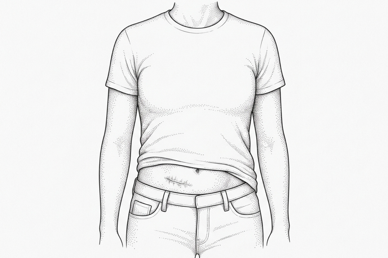
Keloids after abdominoplasty grow beyond the incision line and rarely fade on their own. This guide covers why they form, who is most at risk, and which surgical and non-surgical treatments have evidence behind them.
What causes keloids after a tummy tuck
A keloid tummy tuck scar is a raised, firm, and often discolored scar that grows beyond the boundaries of the original abdominoplasty incision. Unlike a normal surgical scar — which flattens and fades over 12 to 18 months — a keloid continues to expand into surrounding healthy skin and does not resolve on its own.
Here is a quick summary for those seeking immediate clarity:
What it is: An overgrowth of scar tissue driven by excess collagen production, extending past the original incision line.
Who is most at risk:
- People with darker skin tones (Fitzpatrick skin types IV–VI)
- Those aged 10–30 years
- Anyone with a personal or family history of keloid scarring
- Patients with prior wound complications such as infection or seroma
Key treatments, from least to most invasive:
| Treatment | Evidence Level | Best Used For |
|---|---|---|
| Silicone gel sheets | Strong | Prevention and early keloids |
| Compression garments | Moderate | Reducing tension post-surgery |
| Intralesional corticosteroids | Strong | Established, raised keloids |
| Laser therapy | Moderate | Texture, redness, and itch |
| Surgical excision + adjuvant therapy | Strong (combined) | Larger or refractory keloids |
When to seek medical review: If a scar grows beyond the incision line, becomes painful or itchy, or shows signs of infection, consult a qualified healthcare provider promptly.
Abdominoplasty — commonly known as a tummy tuck — is one of the most frequently performed body contouring procedures. It involves a long horizontal incision across the lower abdomen, and in most cases, a separate incision around the navel. Both sites carry some risk of abnormal scarring.
Keloid formation following abdominoplasty is not common — published data place the incidence of keloid and hypertrophic scars after this procedure between 1% and 3.7%. But for the patients it affects, the consequences go well beyond aesthetics. Keloids can cause persistent pain, itching, and significant psychological distress, particularly when they appear across a highly visible area of the body.
What makes keloids especially challenging is their tendency to recur. Surgical removal alone carries a recurrence rate as high as 45–100%, which is why current clinical thinking strongly favors multimodal treatment — combining surgical, pharmacological, and physical approaches.
This guide explains the biology behind keloid formation at tummy tuck incision sites, the evidence behind prevention strategies, and the full range of treatment options available as of July 2026.
This content is for informational purposes only and does not constitute medical advice. Consult a qualified healthcare professional for diagnosis and treatment.
How a keloid tummy tuck scar forms
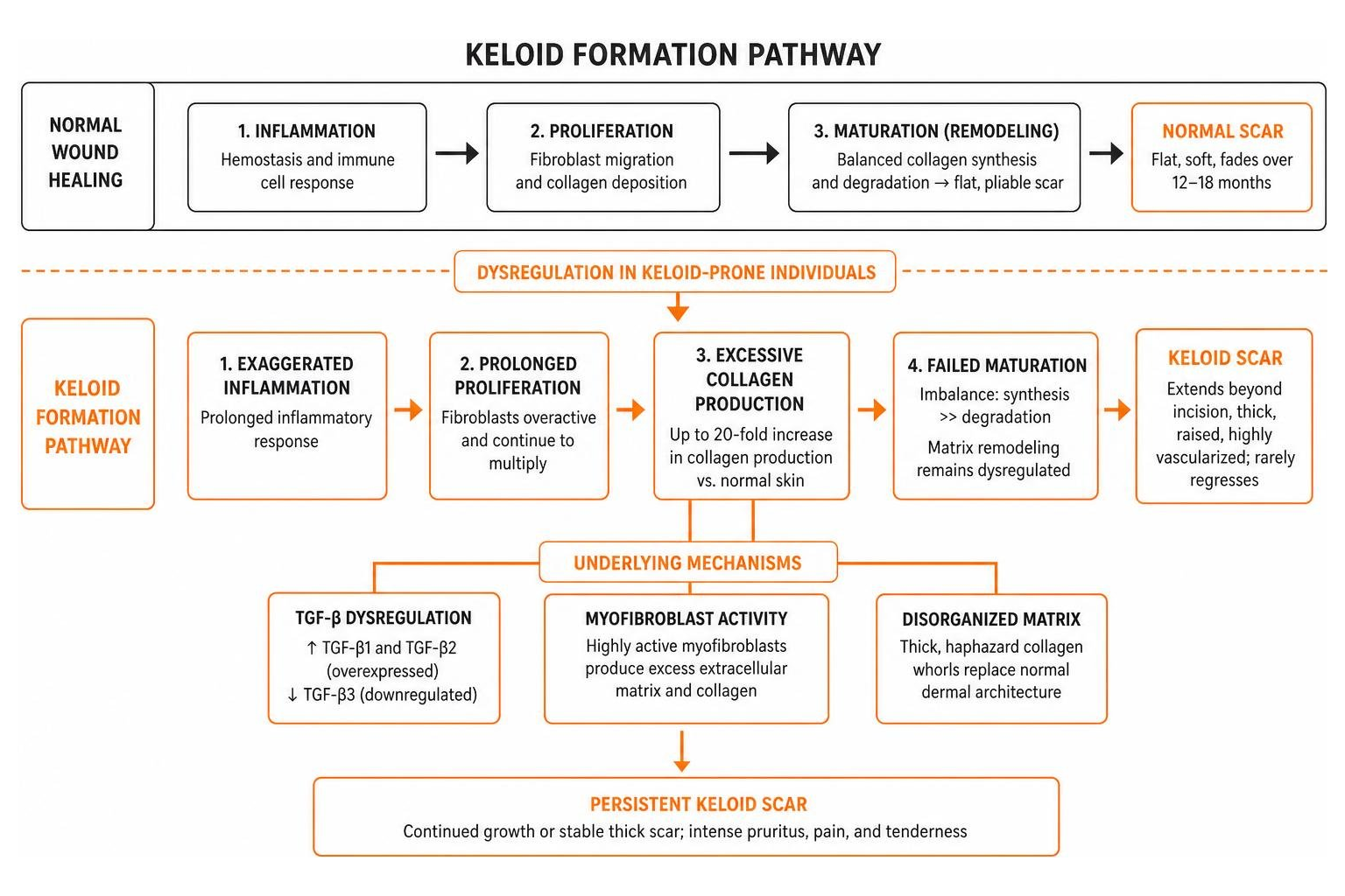
To understand why a keloid tummy tuck scar forms, it is necessary to examine the biological phases of wound healing. Normal wound healing progresses through three distinct stages: inflammation, proliferation, and maturation. During the maturation stage, the body balances the synthesis of new collagen with the degradation of old collagen to remodel the scar tissue into a flat, pliable line.
In keloid-prone individuals, this regulatory mechanism fails. The proliferative phase becomes pathologically prolonged. Fibroblasts — the primary cells responsible for synthesizing the extracellular matrix (ECM) and collagen — continue to multiply and produce collagen at an accelerated rate. Research indicates that keloid tissues exhibit up to a 20-fold increase in collagen production compared to normal healthy skin, whereas hypertrophic scars show only a 3-fold increase.
This dysregulated matrix remodeling is discussed in detail in the Keloid Scar Formation Stages guide. When this occurs along a tummy tuck incision, the scar transitions from a typical post-surgical line into a thick, raised, and highly vascularized nodule.
It is clinically vital to distinguish between normal wound healing, hypertrophic scars, and keloids. While hypertrophic scars are common — occurring in 40% to 70% of surgical patients — they present differently from keloids. The clinical differences are outlined in the comparison table below:
| Clinical Feature | Normal Healing Scar | Hypertrophic Scar | Keloid Scar |
|---|---|---|---|
| Growth Boundaries | Confined strictly to the original incision line. | Confined to the original incision line; does not spread. | Extends beyond the original incision line into healthy tissue. |
| Onset Timing | Fades gradually starting at 3 to 6 months. | Typically appears within 4 to 8 weeks post-surgery. | Can appear months or even over a year post-surgery. |
| Regression | Spontaneously flattens and fades over 12–18 months. | Often regresses spontaneously after 6 to 18 months. | Rarely regresses; continues to grow or remains stable indefinitely. |
| Histology | Organized, parallel collagen bundles. | Fine, wavy, and regularly arranged collagen fibers. | Thick, disorganized, haphazard collagen whorls. |
| Symptoms | Minimal to no physical symptoms after initial healing. | Mild itching or tenderness during the active phase. | Intense pruritus, sharp pain, and physical tenderness. |
For patients seeking further reading on localized hypertrophic reactions, the resource on Tag/Hypertrophic Scars provides additional clinical context.
What a keloid looks like up close
Under microscopic examination, the structural differences of a keloid tummy tuck scar become clear. The normal dermal architecture is replaced by large, thick, closely packed collagen fibers arranged in disorganized, haphazard patterns, whorls, or circular clusters.
This cellular abnormalities are driven by a highly active population of myofibroblasts and a profound dysregulation of the Transforming Growth Factor-beta (TGF-β) signaling pathway. Specifically, there is a marked overexpression of TGF-β1 and TGF-β2, combined with a downregulation of TGF-β3. Because TGF-β1 and TGF-β2 are potent stimulators of collagen synthesis, their uncontrolled activity prevents the scar from reaching a stable state of maturation.
For an in-depth exploration of the cellular mechanisms, genetic pathways, and mechanical triggers that initiate this process, refer to the Keloid Causes Complete Guide.
Who is most likely to get keloids
While local wound factors play a role, systemic and genetic predispositions are the primary determinants of whether a patient will develop a keloid tummy tuck scar.
- Melanin-Rich Skin: Individuals with darker skin tones (Fitzpatrick skin types IV through VI) are more susceptible. The incidence rate of keloids ranges between 6% and 16% in African populations, with a similarly elevated prevalence in Asian and Hispanic populations. This susceptibility is linked to increased melanocyte activity and a more robust cellular response from dermal fibroblasts.
- Age Distribution: Keloids occur most frequently in patients between 10 and 30 years of age. This period of life is characterized by higher hormone levels, elevated skin tension, and a highly active immune response, all of which can drive fibroproliferative disorders.
- Genetic Predisposition: More than 50% of all keloid patients have a documented family history of keloid scarring. This strong hereditary link suggests autosomal dominant or polygenic inheritance patterns.
- Mechanical Tension: The lower abdomen is subject to constant mechanical forces from posture changes, breathing, and core movement. This localized tension acts as a physical trigger, stimulating mechanoreceptors on fibroblasts to upregulate collagen production.
For patients with these risk profiles, preoperative counseling is crucial. Strategies for mitigating these risks are covered in the guides on Abdomen Keloid Scar Reduction and Tag/Keloid Scars.
How surgeons reduce scarring
The prevention of a keloid tummy tuck scar begins in the operating room. A plastic surgeon can employ specific intraoperative techniques to minimize mechanical stress and inflammation, which are the main triggers for abnormal healing.
The most critical factor in scar prevention is tension-free closure. High tension across a healing wound stimulates fibroblasts to deposit dense extracellular matrix. To combat this, surgeons use deep wound closure techniques, such as the Lockwood technique. This approach involves placing high-strength, slowly absorbing sutures into the deep superficial fascial system (SFS). By anchoring the deep tissues, the tension is distributed across the deep abdominal wall rather than the superficial skin layer.
Additionally, progressive tension sutures (PTS) are placed throughout the abdominal flap. These sutures secure the skin flap to the underlying deep fascia, which has two benefits:
- It eliminates dead space, reducing the risk of seroma (which occurs in 5% to 43% of abdominoplasties).
- It prevents the skin edges from pulling apart, reducing surface tension.
Surgical precision is also important during laparoscopic access. While laparoscopy is minimally invasive, port sites can still develop abnormal scars. A retrospective case series published in PMC highlighted that laparoscopically induced keloids represent approximately 1.6% of keloid databases, with a mean onset time of 1.07 ± 0.31 years postoperatively. This highlights that even minor incisions on the abdomen require careful closure and gentle tissue handling to prevent long-term keloid complications.
Belly button incisions and keloid risk
The navel (umbilicus) represents a unique challenge during abdominoplasty. As the only physiological scar on the human body, its reconstruction (umbilicoplasty) requires careful planning. Circular incisions around the umbilicus are highly prone to contracture and keloid formation due to the continuous circumferential tension.
Because of this continuous circumferential tension, surgeons emphasize tension-reducing, non-circular incision designs when reconstructing the umbilicus.
To prevent such complications, modern techniques favor the use of local tissue rearrangements, such as a transposition flap. Rather than a simple circular cut, the surgeon designs a small triangular or rhomboid flap to break up the scar line. This approach is supported by a study in *Archives of Aesthetic Plastic Surgery*, which demonstrated that using a transposition flap to reconstruct the umbilical area preserves the natural depression of the navel, redirects mechanical tension, and significantly reduces the rate of keloid recurrence.
Wound care and non-surgical treatments
Once the surgical closure is complete, the focus shifts to postoperative management. The early weeks following surgery represent a critical window for scar modulation.
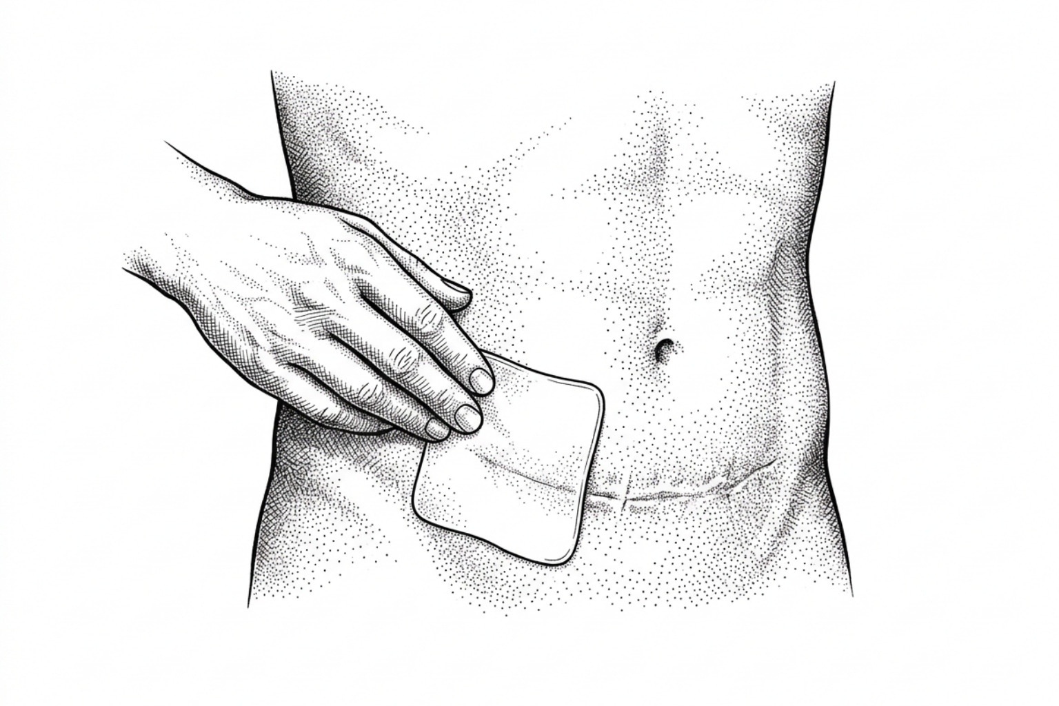
Postoperative care should begin as soon as the epithelial barrier has fully closed (typically 10 to 14 days after surgery). Standard protocols include:
- Compression Therapy: Wearing a medical-grade abdominal binder or compression garment around the clock for at least 14 to 30 days. Compression limits localized edema, reduces capillary perfusion (which starves the hyperactive scar tissue of oxygen and nutrients), and helps align collagen fibers.
- Tension Shielding: Using paper tapes or specialized adhesive strips across the incision to absorb lateral tension forces during early movement.
- Sun Protection: Protecting the healing incision from ultraviolet (UV) radiation for 12 to 18 months. UV light can stimulate melanocytes, leading to persistent post-inflammatory hyperpigmentation.
For a detailed step-by-step recovery timeline, consult the articles on Post Surgery Scar Care and When To Start Scar Treatment After Surgery.
Do silicone sheets and creams work?
Silicone therapy remains the gold standard for non-surgical scar management. It works through two primary mechanisms: occlusion and hydration. By covering the scar, silicone reduces transepidermal water loss (TEWL), signaling the overlying keratinocytes to produce fewer cytokines that stimulate fibroblast activity.
A clinical pilot study published in PMC demonstrated the efficacy of this approach. The study compared conventional 4-mm silicone gel sheets with modified, thinner 2-mm sheets over a 6-month period. Both groups showed significant improvements in scar scale scores compared to untreated controls. Notably, the thinner 2-mm sheets achieved comparable results with zero reported side effects, whereas 20% of patients using the thicker sheets experienced localized itching or skin irritation.
To optimize outcomes, patients are advised to apply silicone sheets for 12 to 24 hours a day for at least 3 to 6 months. For patients who find sheets difficult to secure under clothing, topical silicone gels are a suitable alternative.
For more information on selecting appropriate topical options, refer to the following clinical guides:
Steroid injections and laser therapy
When a scar begins to show early signs of keloid progression — such as thickening, localized redness, or persistent itching — more targeted interventions are required.
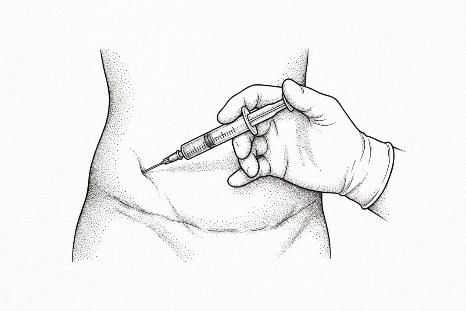
Intralesional Corticosteroid Injections: The most common first-line treatment for active keloids is the injection of triamcinolone acetonide (TAC), typically at concentrations of 10 to 40 mg/mL. Injections are administered directly into the scar tissue every 4 to 6 weeks. Corticosteroids work by suppressing inflammatory mediators, inhibiting fibroblast proliferation, and promoting the breakdown of collagen. While highly effective at flattening scars, potential side effects include localized skin thinning (atrophy), telangiectasia (visible blood vessels), and pigment loss.
Laser Therapy: For vascular and highly symptomatic scars, light-based treatments are often combined with injections.
- Pulsed Dye Laser (PDL - 585/595 nm): Targets hemoglobin in the blood vessels supplying the scar, reducing redness and slowing the scar's growth by limiting its nutrient supply.
- Fractional Ablative Lasers (CO2 or Er:YAG): Create microscopic treatment zones in the scar tissue to help reorganize disorganized collagen fibers and improve pliability.
These non-surgical options are discussed in depth in the resources on Keloid Treatment Without Surgery and Non Invasive Keloid Removal.
Surgery, radiation, and combined treatments
For large, painful, or treatment-resistant keloids, surgical scar revision may be considered. However, simple surgical removal of a keloid is rarely performed alone due to recurrence rates exceeding 50%. Instead, modern protocols combine surgical excision with immediate adjuvant therapies to prevent recurrence.
A study published in *Plastic and Reconstructive Surgery - Global Open* evaluated a strict three-part protocol for treating difficult umbilical keloids:
- Surgical Core Excision: Removing the keloid tissue while preserving as much surrounding healthy skin as possible.
- Postoperative Radiotherapy: Administering low-dose superficial radiation (typically 15 Gy delivered in two fractions over consecutive days) starting within 24 to 72 hours of surgery. This radiation targets and controls the hyperactive fibroblasts.
- Wound Self-Management: Following up with continuous silicone tape application and, if early redness or firmness appears, using high-potency steroid plasters.
This combination protocol achieved a recurrence rate of just 8.8% over a 24-month follow-up period, demonstrating the efficacy of a coordinated, multimodal approach.
Other emerging adjuvant treatments include placing Platelet-Rich Plasma (PRP) into the wound bed immediately after excision to promote organized healing, or using intralesional chemotherapy agents. These advanced approaches are reviewed in the Surgery Scar Treatment Complete Guide and the clinical overview on Keloid Scar After Tummy Tuck | Prevention & Treatment Options.
Frequently asked questions
Navigating postoperative care can be challenging. Below are answers to common clinical questions regarding abdominal scar management, with further details available in the resource section on Tag/Abdominal Scars.
How long does it take for a tummy tuck scar to mature?
The process of scar maturation is a slow biological journey. While the skin incision closes within 10 to 14 days, the underlying collagen remodeling continues for 12 to 18 months.
During the first 1 to 3 months, it is normal for the scar to appear pink, firm, and slightly raised as blood flow to the area remains high. Between 3 and 6 months, the body begins to break down excess collagen, and the blood vessels gradually recede. For a normal scar, this results in a flat, pale, and soft line by the end of the first year.
Because of this timeline, most reconstructive surgeons recommend waiting at least 12 months before considering surgical scar revision, allowing the tissues to fully stabilize.
Are keloid scars more common in patients with darker skin tones?
Yes, clinical evidence shows that individuals with darker skin tones (Fitzpatrick skin types IV–VI) have a higher genetic predisposition to keloid formation. This increased risk is linked to cellular differences in melanin-rich skin:
- Increased Melanocyte Activity: Melanocytes (the cells that produce pigment) interact closely with dermal fibroblasts. Following an injury, this interaction can stimulate fibroblasts to produce more collagen.
- Robust Fibroblast Response: Fibroblasts in darker skin tones tend to be larger, more active, and produce higher levels of collagen in response to inflammatory signals.
- Post-Inflammatory Hyperpigmentation (PIH): Even when a keloid does not form, scars on darker skin are more prone to hyperpigmentation, causing them to appear dark brown or purple. Early use of sun protection and targeted topical therapies can help manage this pigment response.
What are the emerging therapies for keloid prevention?
Medical research is continually exploring new ways to treat and prevent keloids. Some of the most promising emerging therapies include:
- Intralesional 5-Fluorouracil (5-FU): This chemotherapy agent is increasingly used in combination with triamcinolone injections. 5-FU works by blocking DNA synthesis, which helps slow down the rapid multiplication of hyperactive fibroblasts. Clinical studies show that combining 5-FU with steroids yields higher flattening rates and fewer side effects (such as skin thinning) than using steroids alone.
- Bleomycin: Another therapeutic agent that has shown promise in clinical trials for flattening stubborn keloids by inhibiting collagen synthesis.
- Novel Topical Formulations: Researchers are investigating topical treatments designed to block specific growth factor pathways (such as TGF-β receptor inhibitors) or reduce local inflammation at the genetic level. While still undergoing clinical trials, these therapies may offer more targeted, non-invasive options in the future.
Key takeaways
Managing a keloid tummy tuck scar requires a proactive and comprehensive approach. Because keloids are driven by a complex mix of genetics, skin tension, and cellular activity, relying on a single treatment is rarely sufficient. The most successful outcomes are achieved through a combination of early prevention, precise surgical techniques, and consistent postoperative care.
For those planning an abdominoplasty or currently managing a healing incision, early monitoring is key. Recognizing the initial signs of abnormal scarring — such as persistent redness, itching, or thickening that extends beyond the incision line — allows for timely, non-surgical interventions when they are most effective.
To help evaluate your healing progress and receive personalized guidance, consider using the Scar Assessment Tool for a structured evaluation of your scar's characteristics.
Works Cited
- Kwon, H.J., Kim, J.H., Lee, C.R., Choi, J., Moon, S.H., Jun, Y.J., & Oh, D.Y. "Umbilical trocar port site keloid management using a transposition flap after laparoscopic surgery." Archives of Aesthetic Plastic Surgery, vol. 27, no. 4, 2021, pp. 125–131.
- Wang, H.C., Li, Z., Yu, N., Huang, J., & Long, X. "Laparoscopic procedures-induced keloids: A retrospective case series study." International Wound Journal, 2022.
- Kakinuma, T., Kagimoto, M., Kaneko, A., et al. "Prevention and management of hypertrophic scars after laparoscopic surgery using silicone gel sheets: a pilot study." The Journal of International Medical Research, 2022.
- Dohi, T., Kuribayashi, S., Aoki, M., Tosa, M., Akaishi, S., & Ogawa, R. "Combination Therapy Composed of Surgery, Postoperative Radiotherapy, and Wound Self-management for Umbilical Keloids." Plastic and Reconstructive Surgery – Global Open, vol. 8, no. 10, 2020.
- Carswell, L., & Borger, J. "Hypertrophic Scarring and Keloids." StatPearls, NCBI Bookshelf, 2023.
This content is for informational purposes only and does not constitute medical advice. Consult a qualified healthcare professional for diagnosis and treatment.