Why Your Body Overreacts to Tiny Scratches
Keloids form when wound healing won't switch off — collagen keeps building long after a cut has closed. Here's what drives that overreaction, who's most at risk, and the treatments with real evidence behind them.
How keloids form
Keloid causes include any form of skin injury — from a minor piercing or acne breakout to a surgical incision or burn — that triggers an abnormal healing response in genetically susceptible individuals.
Common keloid causes at a glance:
- Skin injuries (acne, piercings, tattoos, burns, cuts, surgical incisions)
- Excess collagen production during wound healing
- Genetic predisposition and family history
- Dark skin tone (Fitzpatrick types III–VI)
- Age between 10 and 30 years
- Hormonal changes during puberty or pregnancy
- Mechanical tension on healing skin (chest, shoulders, upper back)
Most scars form, flatten, and fade. A keloid does the opposite.
Instead of stopping once a wound is closed, the body keeps producing collagen — a structural protein that gives skin its strength — far beyond what the repair requires. The result is a raised, firm growth that extends beyond the original wound boundary and, unlike most scars, does not regress on its own.
This is not simply a cosmetic nuisance. Research indicates that 86% of keloid patients report persistent itching, and 46% experience pain. For many people, the psychological impact — on self-esteem and daily social interactions — is just as significant as the physical symptoms.
Understanding why this overreaction happens is the first step toward managing it effectively.
What is a keloid?
To understand keloid causes, one must first distinguish these growths from other forms of scarring. While both keloids and hypertrophic scars involve an overproduction of collagen, their clinical behavior and biological makeup differ significantly.
A hypertrophic scar is a thickened, raised scar that remains strictly within the boundaries of the original wound. These often develop within weeks of an injury and may naturally regress or flatten over several years. In contrast, a keloid is characterized by its invasive nature; it migrates into healthy, uninjured skin surrounding the initial site.

Keloids vs. hypertrophic scars
| Feature | Keloid Scar | Hypertrophic Scar |
|---|---|---|
| Growth Boundary | Extends beyond the original wound | Stays within wound borders |
| Natural Regression | Rarely regresses; often grows | May flatten over time |
| Collagen Structure | Haphazard, thick bundles | Organized, wavy, parallel |
| Common Sites | Chest, shoulders, earlobes | Any area of injury |
| Genetic Link | Strong familial predisposition | Weak or no genetic link |
| Onset | Months to years after injury | 4 to 8 weeks after injury |
According to research from DermNet, keloids are histologically distinct, featuring large, thick, "glassy" collagen bundles. Understanding these differences is vital for diagnosis, as the Keloid Scar Formation Stages progress differently for each type. While a hypertrophic scar might respond to simple pressure, a keloid often requires a more aggressive, multi-modal approach due to its high recurrence rate.
What causes keloids
The genesis of a keloid is almost always rooted in dermal trauma. When the deep layer of the skin (the dermis) is breached, the body initiates a four-stage healing process: hemostasis, inflammation, proliferation, and remodeling. In keloid-prone skin, the "off switch" for the proliferation phase appears to be broken.
Common triggers identified by the American Academy of Dermatology include:
- Body Art and Modifications: Piercings (especially of the earlobe) and tattoos are among the most frequent keloid causes. Earlobe keloids often form "dumbbell" shapes, growing on both sides of the piercing.
- Inflammatory Skin Conditions: Severe acne, particularly cystic acne on the chest or back, can lead to multiple small keloids. For those dealing with this, a Microneedling Acne Keloid Scars Complete Guide may provide insights into specialized treatment options, though traditional microneedling must be approached with extreme caution in keloid-prone individuals.
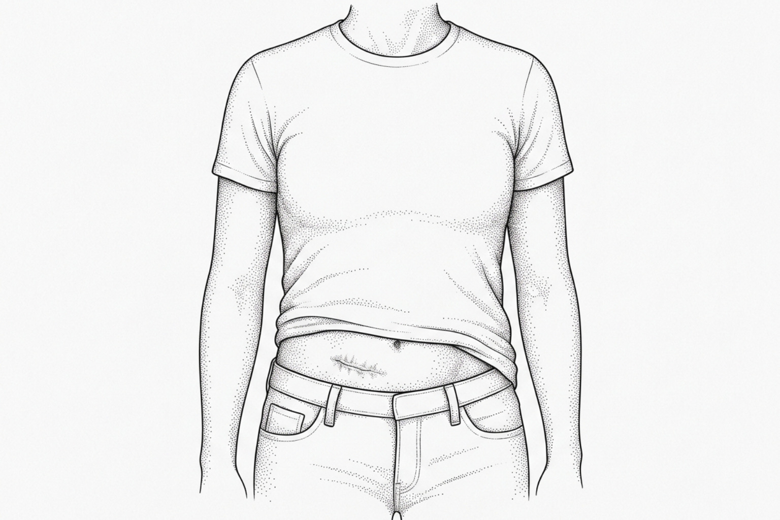
- Surgical Interventions: Surgical incisions, including C-sections and orthopedic surgeries, are significant triggers. The tension of the stitches and the depth of the incision can stimulate excess tissue growth.
- Minor Abrasions: Even seemingly trivial injuries like bug bites, scratches, chickenpox scars, or shaving nicks can serve as keloid causes. In some cases, the original injury was so minor that the patient does not recall it, leading to what are sometimes called "spontaneous" keloids.
What happens at the cellular level
At the cellular level, the primary driver of keloid formation is the fibroblast. Fibroblasts are the cells responsible for synthesizing collagen and the extracellular matrix (ECM). In keloids, these cells are not just more numerous; they are hyperactive and hypersensitive.
Research published in StatPearls highlights the role of Transforming Growth Factor-beta (TGF-β). Specifically, keloid fibroblasts overexpress TGF-β1 and TGF-β2, which are pro-fibrotic signaling proteins. Simultaneously, there is often a decrease in TGF-β3, which typically helps reduce scarring.
This imbalance leads to a metabolic "perfect storm":
- Increased Synthesis: Collagen production in keloids can be up to 20 times higher than in normal skin.
- Decreased Degradation: The body uses enzymes called Matrix Metalloproteinases (MMPs) to break down excess collagen. In keloids, these enzymes are inhibited by high levels of TIMPs (Tissue Inhibitors of Metalloproteinases).
This inability to clear away excess protein results in the dense, fibrous mass characteristic of the condition. For those seeking to address this molecular overactivity, Non Invasive Keloid Removal techniques often focus on modulating these cellular signals without triggering further trauma.
Hormones, genes, and keloid risk
Evidence suggests that hormones play a significant role in the pathophysiology of keloids. The highest incidence occurs between the ages of 10 and 30, coinciding with puberty, when growth hormones and sex steroids are at their peak.
Pregnancy is also known to exacerbate keloid growth or trigger new formations. This is likely due to increased levels of estrogen and progesterone, which can influence inflammatory mediators and increase skin vascularity.
Furthermore, certain rare genetic conditions are associated with a high risk of keloids, such as Rubinstein-Taybi syndrome and Noonan syndrome. For broader information on how these genetic markers manifest, the Tag/Keloid Scars resource provides a library of related clinical observations.
Who's most at risk
While anyone can develop a keloid, certain demographic groups are at a significantly higher risk. This suggests a strong underlying genetic component, even if a single "keloid gene" has not yet been identified.

Skin tone and keloid risk
Clinical data shows that individuals with darker skin tones (Fitzpatrick scales III through VI) are 15 times more likely to develop keloids than those with very light skin. Specifically:
- African Descent: Prevalence rates are self-reported as high as 16% in some Black populations.
- Asian and Hispanic Descent: These groups also show a significantly higher incidence compared to Caucasian populations.
- Family History: Approximately one-third of individuals who develop keloids have a first-degree relative (parent or sibling) with the same tendency.
Yale Medicine notes that keloids are virtually non-existent in albino populations, leading researchers to hypothesize a link between melanocytes (pigment-producing cells) or melanin-related genes and the wound-healing response. Some theories suggest that melanin itself might influence the inflammatory phase of healing, though research is ongoing.
Why skin tension matters
One of the most fascinating aspects of keloid causes is the influence of physical tension on the skin. Keloids do not appear with equal frequency across the body; they favor areas of high mechanical stress.
The "keloid-prone" zones typically include:
- The Presternal Region: The center of the chest is the most common site for keloids, often following heart surgery or even severe acne.
- The Deltoid and Upper Back: These areas experience constant pulling and stretching during arm and torso movement.
- The Earlobes: While earlobes are low-tension areas, they are highly susceptible due to the frequency of piercings and the "trapping" of dermal elements within the piercing tract.
How tension becomes tissue
The process of "mechano-transduction" occurs when skin cells sense physical stretching and convert that mechanical signal into biological activity. When a wound is under tension, fibroblasts are activated to produce more collagen to "strengthen" the area. In keloid-prone individuals, this response is exaggerated.
According to the NCBI Bookshelf, surgical techniques that reduce tension—such as using everted edges or placing incisions along natural skin tension lines (Langer’s lines)—are essential for preventing recurrence.
Treatment options that work
Managing keloids is notoriously difficult because the very act of treating them (especially through surgery) can act as a new trigger. Consequently, a multi-modal approach is the standard of care.
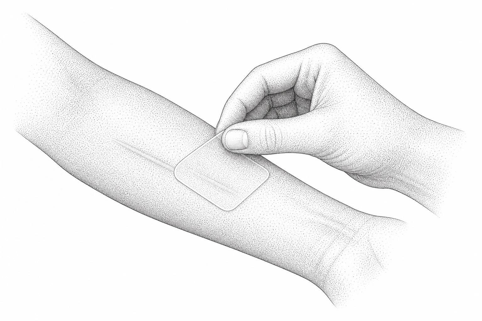
- Silicone Gel and Sheeting: This is often the first line of defense. Evidence suggests that wearing silicone for 12 to 24 hours a day for several months can hydrate the scar and reduce fibroblast activity.
- Pressure Therapy: Especially effective for earlobe keloids, pressure earrings or garments exert constant force (at least 24mmHg) to reduce blood flow and oxygen to the scar tissue, slowing its growth.
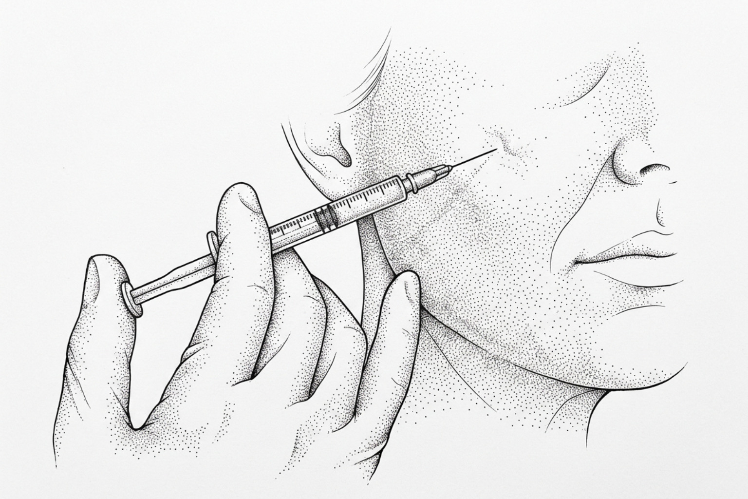
- Corticosteroid Injections: Intralesional triamcinolone acetonide is a common treatment. It works by reducing inflammation and inhibiting the overproduction of collagen. Success rates for flattening vary from 50% to 100%, though recurrence is common.
- Modern Interventions: For stubborn cases, Keloid Treatment Without Surgery or Laser For Keloid Scars can be used to target the vascularity of the scar or break down the dense collagen matrix.
Common questions about keloids
Can keloids form without a visible injury?
Yes. These are often referred to as "spontaneous keloids." However, medical experts believe most "spontaneous" cases are actually the result of minor, unnoticed trauma—such as an ingrown hair, a small pimple, or internal inflammation. MedlinePlus notes that keloids can also appear months or even a full year after an injury has seemingly healed.
Why do keloids cause physical discomfort?
The itching (pruritus) and pain associated with keloids are not just "in the head." Keloid tissue contains a high concentration of mast cells, which release histamine—the same chemical responsible for allergic reactions. Additionally, the rapid expansion of the scar can compress local nerves, leading to a condition known as small nerve fiber neuropathy, which causes sharp or burning pain.
Are keloids a form of skin cancer?
No. Keloids are entirely benign (non-cancerous) growths. They do not spread through the bloodstream to other organs, and they are not contagious. While they involve rapid cell proliferation, it is a localized overgrowth of fibroblasts, not a malignancy. However, because they can occasionally mimic rare skin cancers like dermatofibrosarcoma protuberans, a dermatologist may perform a biopsy if the diagnosis is uncertain.
Key takeaways
The formation of a keloid is a complex biological overreaction where genetics, hormones, and mechanical stress converge. By understanding the primary keloid causes—ranging from molecular signaling imbalances like TGF-β1 overexpression to anatomical high-tension zones—patients and clinicians can better predict and mitigate risk.
While there is no "magic cure" that works for everyone, early intervention is key. Protecting healing wounds from sun exposure, using silicone therapy, and avoiding unnecessary skin trauma are the most effective ways to manage a predisposition to these scars. For a personalized look at your skin's healing patterns, consider a professional Scar Assessment to determine the best path forward.
Works Cited
- American Academy of Dermatology. "Keloid Scars: Causes." American Academy of Dermatology.
- DermNet. "Keloid and Hypertrophic Scar." DermNet NZ.
- Gauglitz, G. G., et al. "Hypertrophic Scarring and Keloids: Pathomechanisms and Current and Emerging Treatment Strategies." Molecular Medicine, 2011.
- Juckett, G., & Hartman-Adams, H. "Management of Keloids and Hypertrophic Scars." American Family Physician, 2009.
- MedlinePlus. "Keloids." U.S. National Library of Medicine Medical Encyclopedia.
- StatPearls. "Hypertrophic Scarring Keloids." NCBI Bookshelf.
- Yale Medicine. "Keloids." Yale Medicine Fact Sheets.
This content is for informational purposes only and does not constitute medical advice. Consult a qualified healthcare professional for diagnosis and treatment.