A Comprehensive Guide to Comparing the Best Filler Scar Treatments
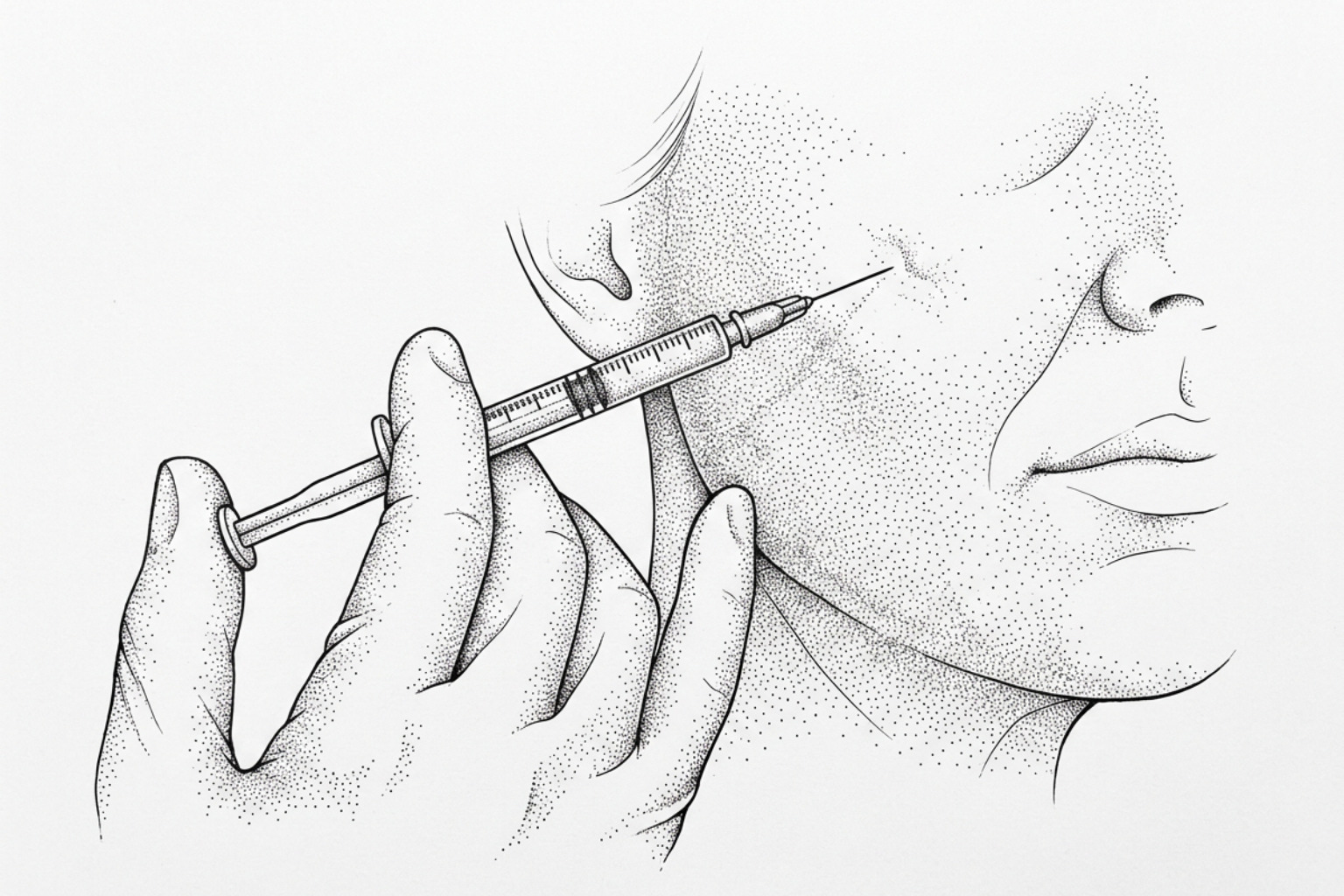
Dermal fillers lift depressed acne scars immediately and, with some materials, stimulate new collagen for years. This guide compares hyaluronic acid, PMMA and biostimulatory options on durability, safety and cost.
How fillers treat acne scars
Finding the best fillers scar treatment depends on scar type, depth, and how long results need to last. Here is a quick reference before diving into the full analysis:
Top dermal filler options for acne scars, compared:
| Filler Type | Best For | Longevity | Reversible? |
|---|---|---|---|
| Hyaluronic acid (HA) | Rolling, boxcar scars | Up to 2 years in scars | Yes |
| PMMA (Bellafill) | Deep atrophic scars | Up to 5 years | No |
| Poly-L-lactic acid (PLLA) | Broad volume loss | 2+ years | No |
| Calcium hydroxylapatite | Deeper atrophic scars | 12–18 months | No |
| Autologous fat transfer | Extensive scarring | Variable | No |
About one in five people who develop acne will go on to form permanent scars. For many, those scars are not just a skin concern — they affect confidence, social comfort, and quality of life.
The most common type is the atrophic scar — a depressed mark left behind when tissue is lost during the healing process. Rolling scars, boxcar scars, and ice pick scars all fall into this category. Unlike raised scars, which involve excess tissue, atrophic scars represent a deficit: the skin surface sits lower than the surrounding skin because collagen and subcutaneous volume were destroyed.
Topical products alone cannot restore that lost volume. That is where injectable fillers come in.
Dermal fillers work by physically lifting the base of a depressed scar and, in many cases, stimulating the body's own collagen production over time. The FDA has approved more than 30 dermal filler formulations, and more than 3.4 million filler injections are performed in the United States every year — a figure that reflects how widely adopted these treatments have become.
But not all fillers work the same way, and not all scars respond to the same approach. The sections below compare the leading options using current clinical evidence, so readers can understand what the research actually supports — and what questions to bring to a qualified clinician.
Glossary for best fillers scar treatment:
Why acne scars leave dents
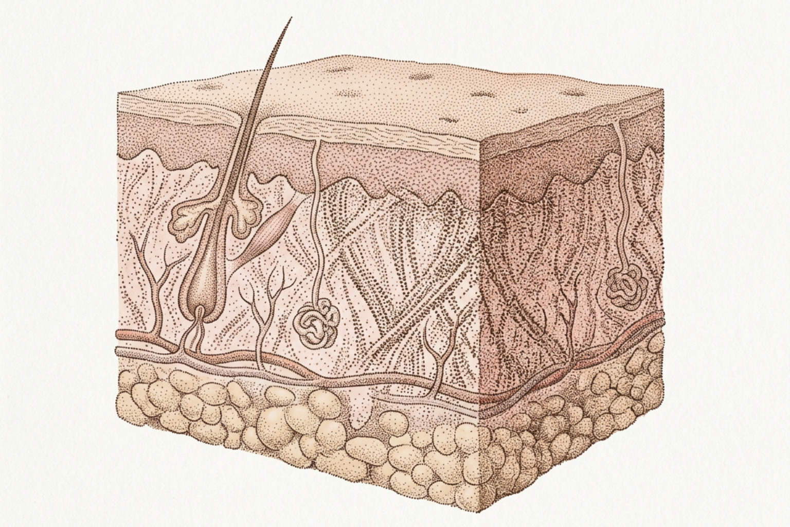
To understand why certain dermal fillers are highly effective for scar revision, it is necessary to examine the underlying biology of scar formation. Atrophic acne scars are the structural consequence of an altered inflammatory response during moderate-to-severe acne outbreaks. When a profound inflammatory lesion occurs in the dermis, the body attempts to repair the damage by producing granulation tissue. However, if the inflammatory process degrades the surrounding extracellular matrix (ECM) faster than the body can synthesize new structural proteins, a net loss of dermal tissue occurs.
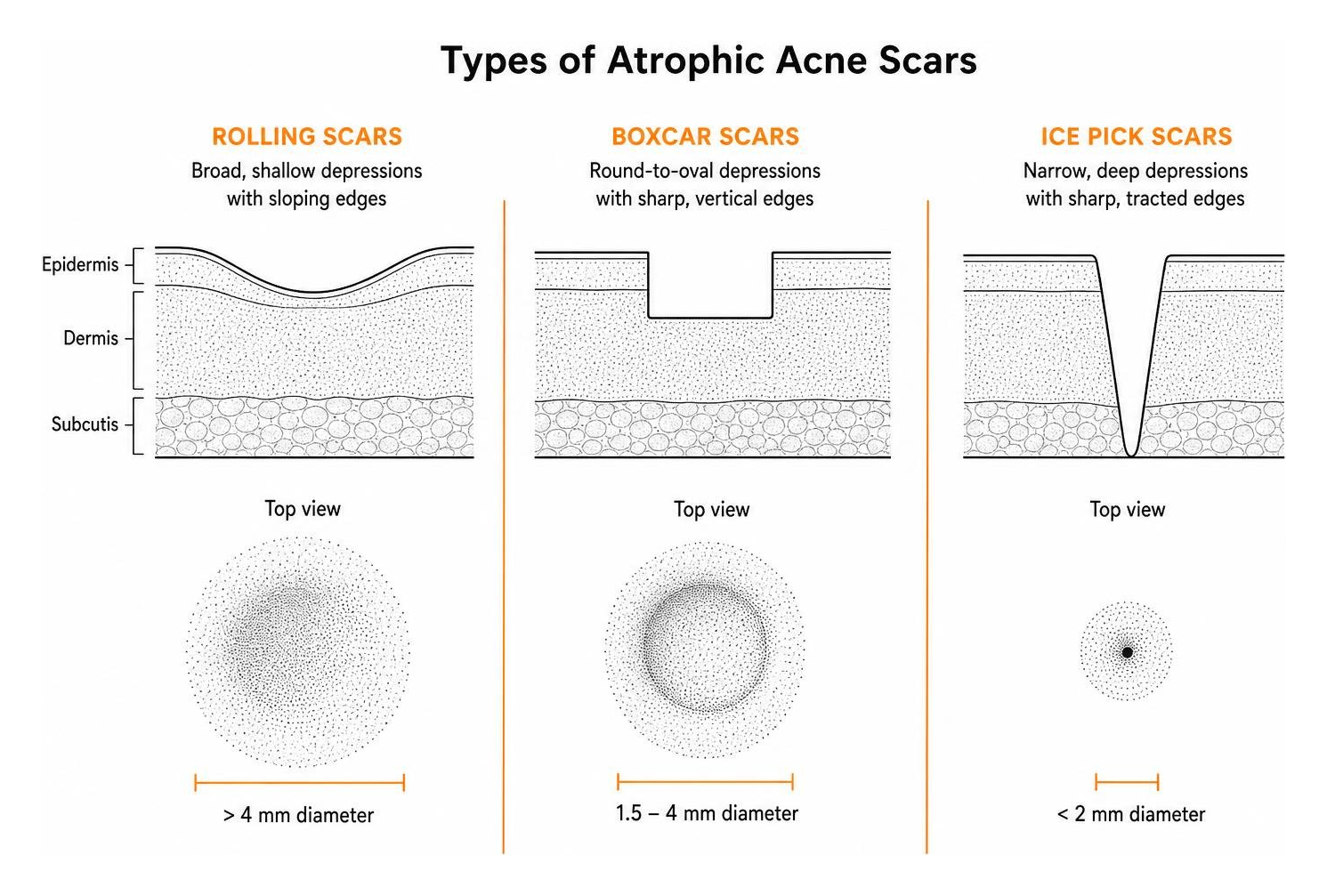
This tissue deficit manifests as three primary types of atrophic scars:
- Rolling Scars: Broad, shallow depressions (typically greater than 4 mm in diameter) that have gently sloping edges. These are frequently anchored to the underlying subcutaneous tissue by fibrous bands.
- Boxcar Scars: Round-to-oval depressions with sharp, vertical margins. They can be shallow or deep, and they occur when the dermal structural support is lost abruptly.
- Ice Pick Scars: Narrow (less than 2 mm), deep, and sharply tracted depressions that extend vertically into the deep dermis or superficial subcutis.
When evaluating Atrophic Scar Filling Treatments, the primary therapeutic objective is to address this volume deficit. Dermal fillers achieve this through two distinct mechanical and biological pathways:
- Immediate Mechanical Lifting: Injecting a cohesive gel directly beneath the depressed scar base provides instantaneous physical support. The filler material occupies the vacant space in the dermal or subdermal layer, pushing the overlying epidermis upward to align with the surrounding skin plane.
- Fibroblast Stimulation and Extracellular Matrix Restoration: The mechanical pressure exerted by the injected filler stretches the surrounding dermal fibroblasts. Research in cell biology indicates that stretching fibroblasts triggers a cascade of intracellular signals that upregulate the synthesis of endogenous Type I and Type III collagen, as well as elastin. Over time, this biostimulatory effect replaces the degrading filler material with the patient's own living tissue, leading to sustained structural improvement.
Types of fillers compared
Selecting the best fillers scar treatment requires a detailed understanding of how different synthetic and autologous biomaterials behave within the skin. The table introduced in the opening section outlines the primary categories, but their biological mechanisms and clinical indications differ substantially.
Hyaluronic acid: results and reversibility
Hyaluronic acid (HA) is a naturally occurring glycosaminoglycan found abundantly within the human extracellular matrix. It is highly hydrophilic, capable of binding up to 1,000 times its weight in water, which allows it to provide immediate, soft volume restoration.
For patients seeking a highly predictable and safe entry point into scar revision, HA is widely considered the gold standard due to its immediate reversibility. If an over-correction, asymmetry, or vascular complication occurs, the enzyme hyalurnidase can be injected to rapidly degrade the HA polymer within hours.
Historically, HA fillers were thought to offer only short-term benefits of 6 to 12 months. However, robust clinical evidence has challenged this timeline. A landmark Prospective Clinical Trial Demonstrating the Efficacy of Hyaluronic Acid Filler for the Improvement of Atrophic Facial Scars up to 2 years demonstrated that a specific cohesive HA filler (VYC-17.5L) maintained significant volume correction for up to 24 months.
This extended longevity in scar tissue, compared to dynamic facial wrinkles, is attributed to the lack of repetitive muscle movement in scarred areas. Without constant mechanical shear stress, the enzymatic and free-radical breakdown of the cross-linked HA is dramatically slowed. Furthermore, as the HA slowly degrades, its micro-particles continue to bind water and stimulate localized fibroblast stretching, maintaining a stable volume.
These findings were corroborated by A split‐face, blind, randomized placebo‐controlled clinical trial investigating the efficacy and safety of hyaluronic acid filler for the correction of atrophic facial scars - PMC. The trial showed that HA filler achieved a 65% reduction in the absolute count of rolling scars compared to just a 15% reduction on the saline-treated control side. This highlights that while the mechanical act of needling provides a minor therapeutic benefit, the presence of the HA polymer is essential for substantial clinical correction. For more clinical context on these applications, refer to the overview on fillers for scar treatment.
Longer-lasting fillers: PMMA and biostimulators
When long-term or semi-permanent correction is the primary objective, biostimulatory fillers are often utilized. Unlike HA, which acts primarily as a passive volume placeholder, biostimulatory agents are designed to trigger a controlled, localized foreign-body response that maximizes de novo collagen synthesis.
- Polymethyl-Methacrylate (PMMA): PMMA microspheres suspended in a bovine collagen carrier (commercially known as Bellafill) represent the only injectable filler formulation with a specific FDA approval for the treatment of moderate-to-severe atrophic acne scars. Upon injection, the temporary bovine collagen provides immediate lift. As the body metabolizes this carrier over several months, the PMMA microspheres remain encapsulated in the dermis, acting as a permanent scaffold that stimulates continuous host collagen deposition. Clinical data shows these results can persist for up to 5 years. However, because PMMA is non-reversible, it carries a higher risk of late-onset granulomas (permanent inflammatory bumps) if placed too superficially.
- Poly-L-Lactic Acid (PLLA): PLLA is a biodegradable, biocompatible synthetic polymer. When injected into the deep dermis or subcutaneous space, it does not provide immediate volume. Instead, it slowly degrades over several weeks, provoking a sub-clinical inflammatory response that recruits macrophages and fibroblasts to deposit new collagen. Results appear gradually over 2 to 3 months and typically last more than 2 years.
- Calcium Hydroxylapatite (CaHA): CaHA microspheres suspended in a carboxymethylcellulose gel provide immediate volume followed by long-term biostimulation. In scar revision, CaHA is often highly diluted (hyper-diluted) to allow for superficial dermal placement without the risk of visible clumping.
While these biostimulatory options provide exceptional longevity, they are not suitable for all scar types. For instance, deep, narrow ice pick scars cannot accommodate the particle size of these fillers without a high risk of superficial nodule formation. Specialized techniques are required when addressing these narrow tracts, as discussed in the guides on dermal fillers for ice pick scars and ice pick scar fillers.
How well fillers work, and what to combine
The clinical success of scar revision is measured using standardized tools such as the Global Aesthetic Improvement Scale (GAIS) and the Goodman and Baron qualitative grading system. In clinical trials evaluating high-concentration HA fillers, up to 92% of treated subjects reported visible improvement on the GAIS and expressed high satisfaction at the 2-year mark. Furthermore, objective 3D skin profilometry (using structured light projection to measure skin topography) has demonstrated an average 20% reduction in the deepness of atrophic scars after 4 months of treatment.
However, a single therapeutic modality is rarely sufficient to address complex, multi-patterned scarring. This is particularly true for tethered scars — rolling or boxcar scars that are bound down to the deep fascia by dense bands of fibrotic tissue.
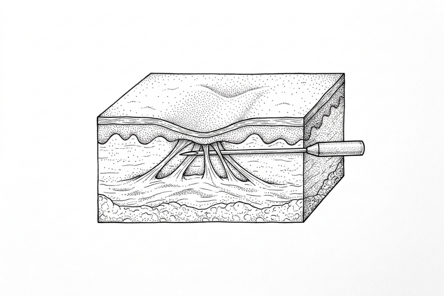
To optimize outcomes, clinicians frequently implement combination protocols that pair fillers with physical release techniques and surface-remodeling devices.
Pairing subcision with fillers
Subcision is a surgical technique wherein a specialized needle or blunt cannula is inserted parallel to the skin surface to physically sever the fibrous tethers pulling the scar downward.
A split-face study published in the Clinical Dermatology Review, titled Hyaluronic Acid Filler versus Biofiller and... : Clinical Dermatology Review, compared commercial HA fillers against autologous biofillers (derived from platelet-poor plasma) combined with platelet-rich plasma (PRP). The study demonstrated that while both modalities significantly reduced scar volume, the mechanical release of the scar base was a critical prerequisite for achieving uniform elevation.
When subcision is performed alone, the severed tissue has a strong tendency to re-tether during the healing phase, pulling the scar back down. Injecting a dermal filler immediately after the subcision procedure serves a vital dual purpose:
- It acts as a physical spacer, preventing the severed fibrotic edges from re-attaching.
- It provides immediate structural support to the newly liberated skin.
For a deeper understanding of these surgical maneuvers and recovery timelines, explore Subcision for Scars, the safety advantages of Blunt Cannula Subcision, and what clinical changes to expect Subcision After One Session.
Combining fillers with lasers and RF
To address both deep volume loss and superficial textural irregularities (such as rough skin texture and enlarged pores), clinicians often combine fillers with fractional CO2 lasers or microneedling. However, the sequencing of these treatments is critical.
Because deep fractional lasers and radiofrequency microneedling devices generate substantial thermal energy in the dermis, they can accelerate the degradation of superficially placed HA fillers. Therefore, the established clinical consensus is to perform all energy-based resurfacing sessions before injecting dermal fillers, or to wait several weeks post-injection to avoid compromising the filler's structural integrity.
What to expect as a patient
Recovery, side effects, and risks
The downtime associated with dermal fillers for scar treatment is minimal, particularly when compared to ablative laser resurfacing. Most patients can resume their normal daily activities immediately following the procedure. However, localized side effects are common and typically resolve within 3 to 7 days:
- Transient Erythema (Redness): Caused by localized inflammation from the needle punctures.
- Mild Edema (Swelling): Particularly common with hydrophilic HA fillers, which draw in water rapidly in the first 48 hours.
- Bruising (Ecchymosis): Can occur if a superficial capillary is disrupted. Applying ice packs with gentle pressure immediately after injection can significantly reduce this risk.
To explore how these recovery profiles fit into a broader, non-surgical treatment plan, refer to the Non-Invasive Acne Scars Complete Guide.
Fillers vs. lasers and chemical peels
Dermal fillers, lasers, and chemical peels target entirely different structural levels of the skin:
- Dermal Fillers: Primarily address volume loss in the deep dermis and subcutaneous space. They are highly effective for rolling and deep boxcar scars but do not alter the surface texture or pigment of the overlying epidermis.
- Laser Resurfacing (Fractional CO2, Er:YAG): Targets the epidermal and upper dermal texture. By creating microscopic zones of thermal damage, lasers force the skin to shed damaged outer layers and remodel superficial collagen. This is excellent for shallow boxcar scars and overall skin tone, but has limited efficacy for deep volume deficits.
- Chemical Peels (such as TCA Cross): Utilize high-strength acids to cause controlled chemical coagulation of the skin. TCA Cross (Trichloroacetic Acid Chemical Reconstruction of Skin Scars) is highly effective for narrow ice pick scars, where the acid destroys the epithelial lining of the scar tract, forcing it to close via secondary intention.
For a comprehensive evaluation of how these modalities compare and how to choose between them, consult What Works for Scars and the Scar Revision Treatment Complete Guide.
Are fillers right for you?
A clinician determines suitability using a simple diagnostic maneuver known as the Dimple Sign Test (or distensibility test).
By applying lateral, inward digital pressure with the fingers on either side of the atrophic scar, the clinician observes how the tissue behaves:
- Negative Dimple Sign (Distensible Scar): If the scar flattens out completely and aligns with the surrounding skin plane when the skin is stretched, it is considered distensible. This indicates that the scar is primarily caused by a localized loss of dermal volume without severe underlying fibrotic anchoring. These scars are excellent candidates for direct dermal filler injection.
- Positive Dimple Sign (Tethered Scar): If the center of the scar remains puckered or dimples further downward when lateral pressure is applied, it is tethered to the deeper structures. This indicates the presence of strong fibrotic bands. These scars are poor candidates for standalone fillers and must undergo subcision prior to filler placement to achieve a successful lift.
Additionally, the clinician must evaluate the epidermal integrity. If the edges of a boxcar scar are highly fibrotic and white (scarred), a filler alone will not soften those hard margins; surface resurfacing will be required.
Key takeaways
Injectable dermal fillers represent a highly effective, clinically validated intervention for correcting the volume deficits associated with atrophic acne scars. Whether utilizing the immediate reversibility and proven 2-year longevity of advanced hyaluronic acid formulations, or the multi-year structural support of biostimulatory agents like PMMA, matching the biomaterial to the specific scar morphology is essential.
Because acne scarring is rarely uniform, the most successful outcomes are achieved through personalized, multi-modality treatment plans. This often involves pairing subcision to release tethered tissue with precisely placed fillers to maintain that elevation, followed by surface resurfacing to refine the skin's texture.
Achieving optimal results requires realistic expectations, an understanding of the underlying anatomy, and a formal clinical evaluation. To begin analyzing your unique skin profile, you can Assess your scars with our interactive tool.
For further scientific reading on wound healing and dermatological interventions, explore our library of clinical topics at Scar Healing Topics or subscribe to our research updates at Scar Healing Subscribe.
Works Cited
- Siperstein, R., Nestor, E., & Meran, S. "Prospective Clinical Trial Demonstrating the Efficacy of Hyaluronic Acid Filler for the Improvement of Atrophic Facial Scars up to 2 Years." Dermatologic Surgery, vol. 50, no. 12, 2024, pp. 1143–1148.
- Siperstein, R., Nestor, E., Meran, S., & Grunebaum, L. "A split-face, blind, randomized placebo-controlled clinical trial investigating the efficacy and safety of hyaluronic acid filler for the correction of atrophic facial scars." Journal of Cosmetic Dermatology, 2022.
- "Hyaluronic Acid Filler versus Biofiller and Platelet-rich Plasma in Atrophic Acne Scars – A Prospective Split-face Study." Clinical Dermatology Review, vol. 10, no. 1, 2026, pp. 29–34.
Disclaimer: This content is for informational purposes only and does not constitute medical advice. Consult a qualified healthcare professional for diagnosis and treatment.