A–Z Guide to Fillers for Scar Treatment
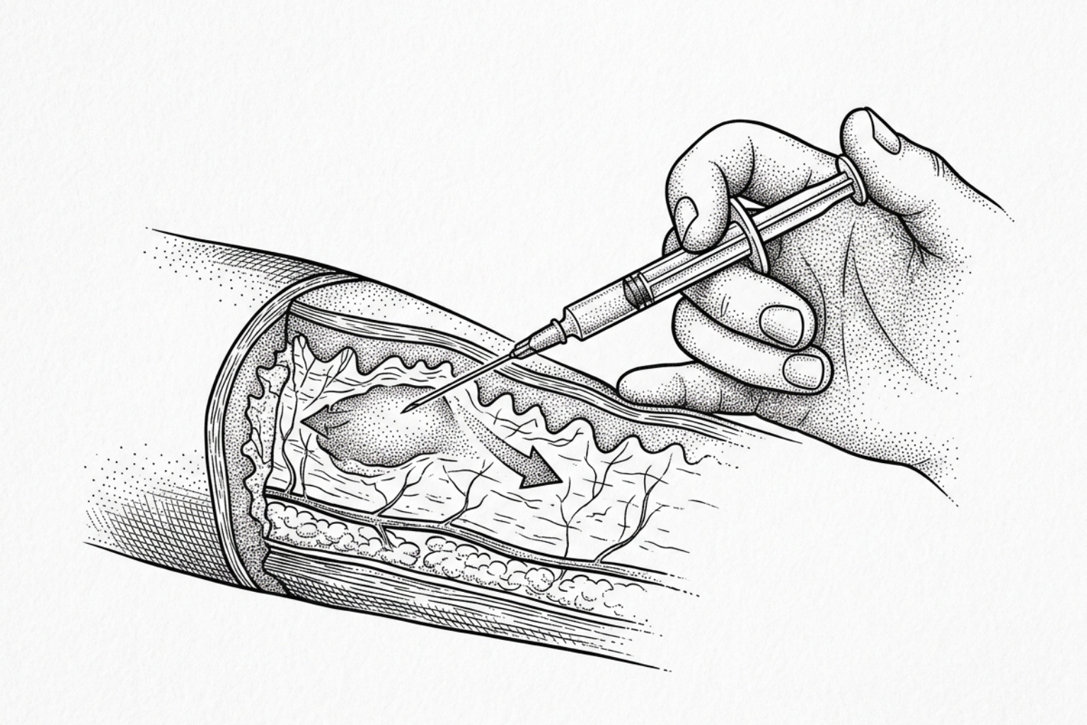
Dermal fillers plump depressed scars almost instantly. Here's how hyaluronic acid, biostimulatory, and permanent fillers compare on results and safety.
How fillers treat scars
Fillers for scar treatment are injectable substances placed beneath depressed skin to restore lost volume, reduce the visible depth of scars, and — depending on the material used — stimulate the body's own collagen production over time.
Here is a quick summary for anyone wanting a fast answer:
| Question | Short Answer |
|---|---|
| What scars respond best? | Atrophic (depressed) scars — especially rolling and boxcar types |
| How do fillers work? | They restore volume under the skin and may trigger new collagen formation |
| How long do results last? | Approximately 6 to 24 months, depending on filler type |
| Is it reversible? | Hyaluronic acid fillers are reversible; most others are not |
| Who should perform it? | A board-certified dermatologist or trained medical professional |
| Main risk? | Transient swelling, bruising; rare but serious: vascular occlusion |
Acne affects roughly 9.4% of the global population, and roughly one in five people who develop acne go on to form permanent scars. For many, those scars are not just a cosmetic concern — they affect self-confidence, social interactions, and quality of life.
Atrophic scars — the sunken, pitted type most commonly left by acne — form when the inflammatory process destroys collagen and fat in the deep layers of the skin. The result is a permanent indentation that sits below the surrounding skin surface, casting a visible shadow.
This is exactly where injectable fillers have found a clinical role.
Unlike topical creams or resurfacing treatments that work on the skin's surface, fillers address the underlying volume deficit directly. Depending on the material injected, they can provide immediate lifting of the scar bed, long-term stimulation of new collagen, or both.
The sections below cover the science, the clinical evidence, the different filler types, and what realistic outcomes look like — so anyone considering this treatment can make an informed decision.
How fillers work
To understand how dermal fillers correct scars, it is necessary to examine the underlying biology of scar tissue. When the skin sustains a deep injury—such as from severe inflammatory acne, trauma, or surgery—the normal architecture of the extracellular matrix is disrupted. The reticular dermis, which accounts for approximately 90% of the skin's thickness, is damaged. During the wound healing process, the skin's inflammatory, proliferative, and maturation phases may fail to synthesize a sufficient volume of collagen and elastin fibers. This localized deficit in structural proteins results in an atrophic, depressed scar.
Injectable fillers for scar treatment work through two primary physiological pathways: immediate mechanical volume restoration and long-term biostimulation.
Instant volume and lift
When a filler is injected into the subcutaneous or deep dermal space directly beneath an atrophic scar, it physically elevates the depressed scar bed. This immediate lifting action aligns the scar with the plane of the surrounding healthy skin, instantly reducing the shadow effects that make depressions visually prominent. This mechanical elevation is particularly effective for distensible scars that can be flattened during a physical stretch test.
How fillers boost collagen
Beyond simple space-filling, many injectable materials act as biological stimulants. The mechanical stretch placed on surrounding dermal tissue by the filler material activates local fibroblasts. Fibroblasts are the primary cells responsible for synthesizing the extracellular matrix, including Type I and Type III collagen.
When these cells are mechanically stretched or chemically stimulated by the injected material, they upregulate the production of endogenous collagen. Over time, as the exogenous filler material slowly undergoes enzymatic degradation or macrophage clearance, it is replaced by a newly synthesized matrix of the patient's own collagen fibers. This biological transition explains why the cosmetic improvements of certain fillers can persist long after the original substance has been metabolized.
Hydration and skin barrier
Healthy skin regeneration relies heavily on maintaining a balanced microenvironment. Research into wound healing has demonstrated that transepidermal water loss (TEWL) plays a regulatory role in tissue remodeling. When the epithelial barrier is compromised or immature, elevated TEWL triggers inflammatory cascades.
For instance, Scientific research on silicone gel occlusion shows that preventing excessive water loss through semi-occlusive barriers downregulates inflammatory cytokines (such as IL-1 and IL-8) and minimizes abnormal collagen deposition. While topically applied silicone gel manages TEWL from the surface to prevent hypertrophic scarring, injectable hyaluronic acid fillers work from within. Hyaluronic acid is highly hydrophilic, capable of binding up to 1,000 times its weight in water. By drawing moisture into the local dermal matrix, it optimizes hydration and supports the cellular signaling necessary for healthy tissue remodeling.
To explore how these biological mechanisms are utilized in clinical practice, readers can access More info about atrophic scar filling treatments.
Types of fillers compared
Clinicians utilize several distinct classes of dermal fillers to treat atrophic scars. These materials vary significantly in their chemical composition, mechanical properties, longevity, and physiological mechanisms.
A comprehensive overview of these differences is detailed in the comparative table below:
| Filler Type | Primary Mechanism | Longevity | Best Suited For | Reversibility |
|---|---|---|---|---|
| Hyaluronic Acid (HA) | Immediate hydrophilic volumization; mild fibroblast stimulation | 6 to 12 months (can persist up to 24 months in scars) | Rolling scars; superficial boxcar scars; first-time patients | Fully reversible with hyaluronidase |
| Calcium Hydroxylapatite (CaHA) | Immediate volume followed by moderate neocollagenesis | 12 to 18 months | Medium-depth atrophic scars; rolling scars | Non-reversible; must degrade naturally |
| Poly-L-Lactic Acid (PLLA) | Gradual, progressive biostimulation of endogenous collagen | 12 to 24+ months | Widespread atrophic scarring; rolling scars; volume loss | Non-reversible |
| Polymethyl-methacrylate (PMMA) | Permanent structural support via microspheres; bovine collagen carrier | Permanent (microspheres remain indefinitely) | Distensible rolling scars; moderate-to-severe acne scars | Non-reversible; surgical excision required for removal |
| Autologous Fat | Living tissue graft; immediate volume and adipose-derived stem cell transfer | Variable (approx. 50% graft survival; surviving fat is permanent) | Large, deep atrophic depressions; post-traumatic/surgical scars | Non-reversible; requires surgical revision if overfilled |
| Biofillers (PPP Gel) | Autologous heated protein gel; physical scaffolding | 3 to 6 months | Widespread atrophic acne scars; patients seeking autologous options | Non-reversible; rapid natural resorption |
A deeper look into how these materials are systematically evaluated in clinical settings can be found in the Systematic review of injectable fillers.
Hyaluronic acid fillers
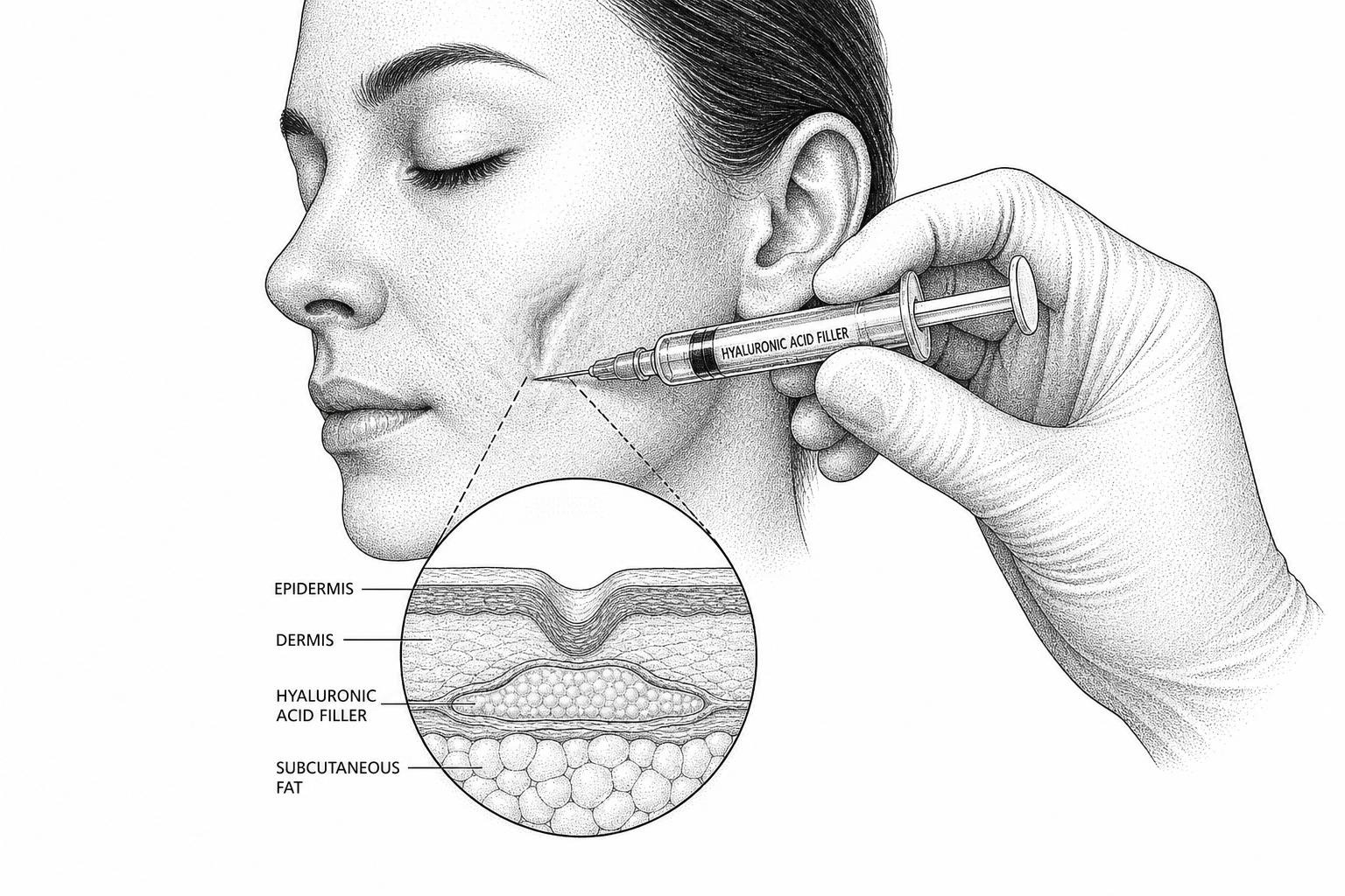
Hyaluronic acid (HA) remains the most widely used injectable material for scar revision. Because HA is a naturally occurring glycosaminoglycan found throughout human connective tissue, it carries an exceptionally low risk of immunogenic reactions.
In the context of scar treatment, HA fillers offer immediate, highly predictable volume correction. The gel is injected directly into the mid-to-deep dermis beneath the scar. Because HA is highly malleable and transparent, it carries a low risk of visible clumping.
A key clinical advantage of HA is its reversibility. If a patient experiences an adverse event, asymmetry, or is unsatisfied with the aesthetic outcome, the enzyme hyaluronidase can be injected to rapidly dissolve the filler.
Clinical studies have demonstrated that HA fillers perform exceptionally well for rolling scars—atrophic depressions with sloped edges that blend gradually into normal skin. To determine if an atrophic scar will respond well to HA monotherapy, clinicians often perform the "Dimple Sign" or stretch test. If stretching the skin adjacent to the scar flattens the depression, the scar is considered distensible and is highly likely to respond favorably to HA injection.
The long-term durability of HA in scar tissue is notably superior to its longevity when injected into dynamic facial wrinkles. A landmark Prospective clinical trial on hyaluronic acid fillers demonstrated that the clinical improvement of atrophic facial scars was sustained for up to two years post-injection without additional touch-ups.
Researchers hypothesize that because scars are static and lack the constant muscular shear forces found in areas like the nasolabial folds or lips, the mechanical breakdown of the cross-linked HA gel is significantly delayed.
Longer-lasting and permanent fillers
For patients seeking longer-lasting or permanent correction, biostimulatory and synthetic permanent fillers are viable clinical options. These materials do not rely solely on the physical volume of the injected gel; instead, they trigger a controlled sub-clinical inflammatory response that recruits macrophages and fibroblasts to synthesize new collagen.
- Poly-L-Lactic Acid (PLLA): PLLA is a biodegradable, biocompatible synthetic polymer. When injected, it does not provide immediate volume. Instead, the microparticles stimulate a gradual foreign-body response over several weeks, leading to progressive neocollagenesis. PLLA is highly effective for treating widespread, diffuse rolling acne scars across the cheeks and temples.
- Calcium Hydroxylapatite (CaHA): Consisting of synthetic calcium hydroxylapatite microspheres suspended in a carboxymethylcellulose gel carrier, CaHA provides immediate physical volume followed by long-term collagen stimulation. Because CaHA is a thick, high-viscosity filler, it must be injected with precision to prevent the formation of visible nodules, particularly in thin-skinned areas.
- Polymethyl-methacrylate (PMMA): PMMA is a permanent synthetic material. In clinical practice, PMMA microspheres are suspended in a carrier gel of bovine collagen. The bovine collagen provides immediate volumetric correction, which gradually degrades over several months. As the carrier gel resorbs, the permanent PMMA microspheres remain in place, acting as a structural scaffold that stimulates the body to encapsulate each microsphere in endogenous collagen. PMMA is the only filler material with specific regulatory approval for the treatment of moderate-to-severe atrophic acne scars.
To understand the histological impact of these materials, clinicians can consult the Literature review on tissue biostimulators. Additionally, for patients presenting with deep, narrow depressions, exploring More info about dermal fillers for ice pick scars provides essential context on the limitations of biostimulatory fillers in highly fibrotic scar subtypes.
Your own tissue and plasma fillers
An emerging alternative to synthetic dermal fillers is the use of autologous biofillers derived from the patient's own blood. Platelet-poor plasma (PPP) gel is prepared by drawing a small volume of the patient's blood, centrifuging it to separate the cellular components, and isolating the plasma fraction that is low in platelets.
This isolated plasma is then subjected to a precise thermal protocol: heating at 80–100°C for approximately 5 minutes to denature and coagulate the plasma proteins, followed by rapid cooling at 0–6°C for 5 minutes. This process transforms the liquid plasma into a cohesive, injectable gel with a semisolid consistency.
Because PPP gel is entirely autologous, it carries zero risk of immunogenic reactions, allergic responses, or foreign-body granulomas. A prospective Clinical study on autologous biofillers evaluated the efficacy of PPP gel as a monotherapy for atrophic acne scars.
The study demonstrated significant improvements in quantitative scar scales over a six-month treatment course of monthly injections, showing immediate volumetric correction and a favorable safety profile. However, because the protein scaffold of PPP gel is naturally degraded by the body's proteolytic enzymes, its longevity is shorter than that of synthetic fillers, typically requiring repeat treatments every few months to maintain the clinical outcome.
Do fillers work, and who they're for
Achieving an optimal clinical outcome with fillers for scar treatment requires precise patient selection and accurate scar classification. Atrophic scars are classified into three primary morphological subtypes:
- Rolling Scars: These are wide depressions (typically >4 mm) with gently sloping edges. They are caused by fibrous anchoring bands that tether the dermis to the underlying subcutaneous tissue. Rolling scars are highly distensible and respond exceptionally well to dermal fillers, as the volume lifts the depression easily once any underlying fibrous tethers are released.
- Boxcar Scars: These are round-to-oval depressions with sharp, vertical borders. Superficial boxcar scars (<0.5 mm depth) can be successfully treated with fillers, whereas deep boxcar scars (>0.5 mm) are highly fibrotic and often require combination resurfacing therapies.
- Ice Pick Scars: These are narrow (<2 mm), deep, sharply demarcated tracts that extend into the deep dermis or subcutaneous layer. Due to their narrow diameter and severe vertical fibrosis, ice pick scars do not respond well to dermal fillers. Attempting to inject filler directly beneath an ice pick scar often causes the material to migrate to the surrounding healthy tissue, worsening the appearance of the depression.
Before proceeding with treatment, clinicians must perform a skin stretch test. If a scar does not flatten when the surrounding skin is gently stretched, it indicates deep dermal tethering. In such cases, dermal fillers should not be used as a monotherapy, as the filler material will simply accumulate around the tethered point, creating a ring of elevation with a persistent central depression.
Patient age and skin type also influence the selection of the filler material. Older patients with age-related dermal atrophy may require biostimulatory fillers (such as PLLA or CaHA) to address generalized volume loss alongside specific scar depressions.
When treating patients with darker skin phenotypes (Fitzpatrick skin types IV–VI), clinicians must exercise caution with highly inflammatory biostimulatory fillers, as any localized sub-clinical inflammation can occasionally trigger post-inflammatory hyperpigmentation (PIH).
For a complete overview of non-surgical scar revision, readers can refer to the More info about non-invasive acne scars complete guide.
Combining fillers with other treatments
In clinical dermatology, monotherapy with dermal fillers is rarely sufficient for complex, multi-textured scarring. To achieve optimal results, fillers are frequently integrated into multi-modality treatment protocols.
Combining mechanical volume restoration with energy-based devices, mechanical resurfacing, or chemical peels addresses both the depth deficit and the surface texture irregularities of the scar tissue.
Combining subcision with fillers
Subcision is a minor surgical procedure where a specialized tri-beveled needle or cannula is inserted beneath the skin to physically sever the fibrotic bands pulling the scar bed downward. Performing subcision immediately prior to injecting a dermal filler is highly synergistic.
Severing the tethers creates a localized pocket that allows the filler material to distribute evenly beneath the scar without resistance. The filler acts as a physical spacer, preventing the severed fibrotic bands from reattaching during the early phases of wound healing.
Laser resurfacing and microneedling
Ablative and non-ablative fractional lasers, along with microneedling, create controlled micro-zones of thermal or mechanical injury in the epidermis and upper dermis. This injury triggers a robust wound healing cascade, leading to epidermal turnover and neocollagenesis.
When combining these modalities with fillers, timing is critical. Energy-based devices that generate deep dermal heat can accelerate the degradation of superficially placed hyaluronic acid or autologous biofillers.
Therefore, clinical guidelines recommend performing laser resurfacing or microneedling sessions first to address surface texture, allowing the skin to heal completely before injecting dermal fillers to correct remaining volume deficits.
Platelet-Rich Plasma (PRP) and Chemical Peels
Platelet-rich plasma (PRP) therapy utilizes the patient’s concentrated platelets, which release high concentrations of growth factors (such as PDGF, TGF-beta, and VEGF) to accelerate tissue repair. Combining PRP with biostimulatory fillers or microneedling enhances the rate of neocollagenesis and reduces post-treatment downtime.
Similarly, focal chemical reconstruction of skin scars (using high-concentration trichloroacetic acid, known as the TCA CROSS method) can be used to treat narrow ice pick scars, while surrounding rolling scars are addressed with dermal fillers.
Benefits of Combination Therapy
- Comprehensive Correction: Addresses both deep volume loss and superficial textural irregularities simultaneously.
- Enhanced Neocollagenesis: Combining physical biostimulation (fillers) with thermal injury (lasers) or growth factors (PRP) produces a larger volume of new collagen than either treatment alone.
- Reduced Risk of Migration: Releasing fibrotic tethers via subcision ensures the injected filler remains positioned directly beneath the scar.
- Improved Patient Satisfaction: Patients experience a more uniform improvement in skin smoothness, pore size, and overall light reflection.
For further clinical insights on identifying and managing different acne scar profiles within a combination treatment plan, readers can visit the More info about acne scars resource page.
Safety, side effects, and risks
While dermal fillers are minimally invasive, they are medical procedures that carry inherent risks and potential complications. Understanding these safety profiles is essential for minimizing adverse events.
Common short-term side effects
Most patients undergo filler injections with minimal disruption to their daily routines. Typical immediate side effects are localized and self-limiting, resolving within 2 to 7 days:
- Transient Erythema: Redness at the injection sites due to localized vasodilation.
- Edema: Mild swelling caused by the physical volume of the filler and the hydrophilic nature of materials like hyaluronic acid.
- Ecchymosis: Minor bruising resulting from localized capillary trauma during needle or cannula insertion.
Rare but serious complications
Though rare, more serious complications can occur and require immediate clinical intervention:
- Nodule and Granuloma Formation: Non-inflammatory nodules can occur due to improper filler placement (e.g., injecting a high-viscosity filler too superficially) or filler clumping. Inflammatory granulomas represent a delayed immune response to the foreign material and may require intralesional corticosteroid injections or surgical excision.
- Vascular Occlusion: The most severe complication of dermal filler injections occurs when filler material is accidentally injected directly into a cutaneous blood vessel, or when excessive external volume compresses a vessel, compromising local blood flow. If left untreated, vascular occlusion can lead to localized skin necrosis, scarring, or in extremely rare cases involving facial arteries, visual impairment. Clinicians mitigate this risk by using blunt-tip cannulas, injecting slowly with low pressure, and maintaining immediate access to hyaluronidase to dissolve HA fillers if ischemic changes (such as skin blanching or severe pain) occur.
When to avoid fillers
Dermal fillers should not be administered to individuals with the following clinical presentations:
- Active Acne or Localized Infection: Injecting through active acne lesions or infected skin can introduce bacteria into the deep dermal space, leading to cellulitis or biofilm formation.
- Known Hypersensitivity: Patients with a history of severe allergies or specific sensitivities (such as bovine collagen allergies for PMMA carriers) must undergo skin patch testing or avoid those materials entirely.
- Systemic Autoimmune or Bleeding Disorders: Active systemic autoimmune diseases can increase the risk of delayed-onset granulomas, while bleeding disorders or concurrent anticoagulant therapy significantly increases the risk of severe hematoma and ecchymosis.
Fillers vs. traditional dressings
Unlike traditional passive scar treatments — such as silicone gel sheets, paper tapes, or polyurethane films which work externally to control hydration and mechanical tension — dermal fillers are active biological therapies. Passive dressings are highly effective at preventing hypertrophic and keloid scars during the early stages of wound healing, as discussed in More info about acne scar treatment natural home remedies.
In contrast, fillers are designed strictly to correct established, mature atrophic depressions. They do not prevent hypertrophic scar formation and should never be injected into active keloids, as this can trigger further pathological tissue proliferation.
Frequently asked questions
How long do dermal fillers last when used for scar correction?
The longevity of dermal fillers for scar correction typically ranges from 6 to 24 months. The exact duration depends on the specific filler material used, the depth of placement, and individual metabolic rates. Hyaluronic acid fillers, which normally last 6 to 12 months in highly mobile areas of the face, often persist for up to 2 years when injected into static scar tissue.
Biostimulatory fillers like PLLA and CaHA generally provide clinical benefits for 12 to 24 months due to the deposition of new endogenous collagen. PMMA is considered a permanent filler, as the synthetic microspheres are not degraded by the body, although the natural aging process and surrounding tissue changes will still affect the long-term aesthetic outcome over several years.
What is the typical recovery time and downtime after the procedure?
The downtime associated with dermal fillers for scar treatment is minimal. Most patients can resume their normal daily routines, including work and light physical activities, immediately following the procedure.
Localized redness, mild swelling, and minor bruising at the injection sites are common but typically resolve within a few days. Patients are generally advised to avoid strenuous exercise, excessive heat exposure (such as saunas), and rubbing the treated areas for 24 to 48 hours post-injection to prevent filler displacement.
Can dermal fillers completely eliminate deep ice pick scars?
No, dermal fillers cannot completely eliminate deep ice pick scars. Ice pick scars are narrow, deep, and highly fibrotic tracts that extend vertically into the deep dermis or subcutaneous tissue. Because the scar is tightly held down by dense collagen bundles, injecting a dermal filler beneath it cannot lift the depression.
Instead, the filler material tends to migrate laterally into the softer, surrounding healthy tissue, which can elevate the edges and make the central depression appear deeper. Deep ice pick scars are best managed using alternative dermatological procedures, such as the TCA CROSS technique, punch excision, or punch elevation, before considering fillers for any remaining superficial contour irregularities.
Key takeaways
Injectable fillers for scar treatment represent a clinically proven, minimally invasive approach to correcting atrophic dermal depressions. By physically restoring lost volume and stimulating endogenous neocollagenesis, these materials address the structural deficits left by inflammatory acne, trauma, and surgical wounds.
While hyaluronic acid remains the clinical standard due to its immediate results and reversibility, biostimulatory and autologous options offer diverse pathways to long-term tissue remodeling.
Despite these advancements, several gaps remain in the scientific literature. There is a need for more long-term, head-to-head randomized controlled trials comparing the long-term safety and efficacy of different filler materials specifically within scar tissue.
Furthermore, establishing standardized objective outcome measures — combining 3D volumetric imaging with patient-reported quality-of-life scales — will help refine clinical algorithms and improve treatment predictability.
Because every scar profile is unique, successful revision requires a personalized approach. Individuals seeking to understand their specific scar characteristics and explore appropriate treatment paths can Get a personalized scar assessment to begin their clinical evaluation.
Works Cited
- "Silicone Gel for Scar Prevention." Chapter 23, Textbook on Scar Management: State of the Art Management and Emerging Technologies. NCBI Bookshelf.
- "The Impact of Tissue Biostimulators on Patients with Post-Operative Scars: A Literature Review." Research, Society and Development, 2025.
- "Monotherapy of Biofiller for Atrophic Acne Scars: A Prospective Nonrandomized Study." PubMed.
- "Injectable Fillers for Atrophic Acne Scars: A Systematic Review of Mechanisms, Evidence, and Clinical Algorithms." Clinical, Cosmetic and Investigational Dermatology.
- "Prospective Clinical Trial Demonstrating the Efficacy of Hyaluronic Acid Filler for the Improvement of Atrophic Facial Scars up to 2 Years." Dermatologic Surgery, 2024.
Medical Disclaimer
This content is for informational purposes only and does not constitute medical advice. Consult a qualified healthcare professional for diagnosis and treatment.