A Comprehensive Guide to Side Effects of Steroid Injections Into Scars
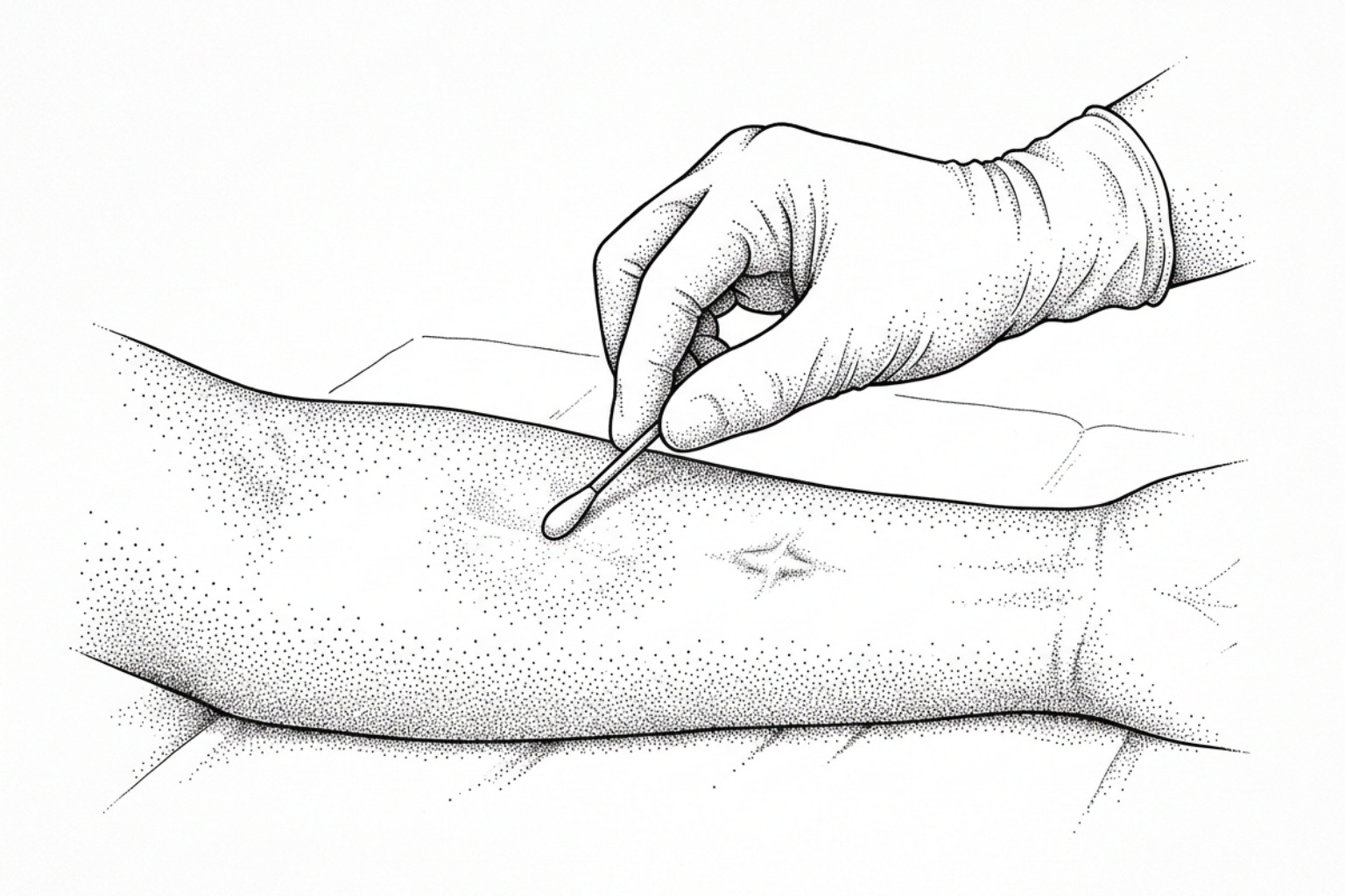
Steroid injections are the most common keloid treatment, and skin thinning, pigment loss and visible veins are their most common costs. Here is how often each happens and what reduces the risk.
How steroid injections affect scar tissue
The most common side effects of steroid injections into scars include localized dermal and subcutaneous skin atrophy (denting or thinning), hypopigmentation (lightening of skin tone), telangiectasia (visible spider veins), and insoluble steroid chalk deposits. While intralesional corticosteroid therapies effectively flatten raised lesions, these adverse reactions occur because corticosteroids alter cutaneous biology and suppress collagen synthesis in both abnormal scar tissue and surrounding healthy skin.
When skin sustains a deep injury extending into the reticular dermis, fibroblasts migrate to the site to lay down an extracellular matrix composed primarily of type I and type III collagen. In standard healing, this process resolves into an organized matrix. However, an understanding of how scars form shows that excessive fibroblast activity, prolonged inflammation, and impaired matrix turnover lead to the dense, raised collagen bundles characteristic of hypertrophic and keloid scars.
As detailed in the Textbook on Scar Management, corticosteroids diffuse across cell membranes and bind to cytoplasmic glucocorticoid receptors. This binding inhibits pro-inflammatory transcription factors, including nuclear factor kappa B (NF-kB) and activator protein 1 (AP-1). This cellular cascade leads to several targeted clinical effects:
- Suppression of Cytokines and Fibroblasts: Corticosteroids decrease transforming growth factor-beta-1 (TGF-beta-1) and vascular endothelial growth factor (VEGF). This halts rapid fibroblast proliferation and promotes fibroblast apoptosis, slowing runaway collagen synthesis.
- Upregulation of Collagenase: Rather than solely stopping collagen production, corticosteroids inhibit alpha-2-macroglobulin and alpha-1-antitrypsin, which are natural circulating inhibitors of collagenase. By neutralizing these inhibitors, the active enzyme collagenase can degrade excess extracellular collagen fibers.
- Symptom Relief and Vasoconstriction: Corticosteroids prompt localized microvascular constriction, depriving rapidly dividing scar tissue of oxygen and nutrients. By suppressing localized histamine and inflammatory mediators, this pathway also directly addresses why scars itch and throb.
While this mechanism reduces pathological scar volume, it is not entirely cell-specific. The same mechanisms that degrade excess collagen in a scar can affect surrounding healthy skin, leading to thinning of the dermis and alterations in local blood vessels.
Side effects at the injection site
When evaluating intralesional corticosteroids as a treatment for keloid scars and hypertrophic lesions, clinical trials indicate an overall positive response rate of 70% to 80%. However, localized adverse effects are common. Systematic reviews and clinical studies report that local adverse reactions occur in approximately 33% to 63% of patients undergoing intralesional corticosteroid therapy.
The primary driver of these side effects is drug extravasation (leakage) into surrounding healthy tissue or subcutaneous layers, excessive concentration, or high injection frequency. When insoluble steroid suspensions remain active in non-scarred tissue, normal collagen turnover is suppressed, leading to localized tissue fragility.
Skin thinning and dents
Dermal and subcutaneous tissue atrophy is one of the most widely documented complications of intralesional triamcinolone. Dermal atrophy manifests as a noticeable indentation, depression, or step-off defect where the skin appears crinkled, translucent, and easily folded.
According to the British Association of Dermatologists intralesional steroid therapy guidelines, this phenomenon happens when the drug suppresses the normal synthesis of extracellular glycosaminoglycans and reduces adipose volume.
The primary factors influencing the severity of skin atrophy include:
- Injection Plane Errors: Placing the needle tip below the dense scar tissue into the subcutaneous fat layer can trigger lipoatrophy—the shrinkage of underlying fat lobules—which causes localized surface dimpling.
- Perilesional Leakage: High injection pressures into hard, fibrotic tissue can cause the suspension to track backward along the needle path, settling in normal adjacent skin.
- Linear Lymphatic Transport: Macromolecules and insoluble steroid crystals can enter the superficial cutaneous lymphatic network. Case reports demonstrate that this can lead to radiating, linear bands of depigmentation and dermal thinning extending outward from the primary scar site.
Colour changes, visible veins and white deposits
Pigmentary and vascular irregularities are additional localized complications seen in clinical dermatology:
- Hypopigmentation and Hyperpigmentation: Hypopigmentation (loss of skin color) occurs because corticosteroids temporarily inhibit melanocyte function and decrease melanin synthesis without completely destroying the melanocytes themselves. This lightening is more noticeable in individuals with darker skin phototypes (Fitzpatrick skin types IV through VI). Hyperpigmentation can also occur as a post-inflammatory response to the needle puncture.
- Telangiectasia: Corticosteroids trigger the release of local angiogenic factors alongside chronic microvascular dilation. This produces visible, branching spider veins across or around the scar border. Unlike transient redness, steroid-induced telangiectasias rarely resolve on their own and may require vascular laser treatment.
- Steroid Chalk Deposits: Suspensions of triamcinolone contain microfine crystalline particles. If injected too superficially into the upper papillary dermis, or if the suspension fails to resuspend properly before administration, these crystals can coalesce. This forms visible, firm white-to-yellow deposits under the skin known as "steroid chalk" or foreign body granulomas.
As outlined in NHS patient guidance on triamcinolone acetonide injections, patients should be informed about these localized cosmetic risks prior to initiating a multi-session regimen.
Whole-body effects and who is most at risk
Because intralesional injections deliver small volumes directly into fibrous tissue, systemic absorption is typically low. However, synthetic corticosteroids enter the general circulation in small amounts. When high drug concentrations, large volumes, or frequent appointments are used, systemic complications can develop. Understanding these biological variables is a core part of reviewing a keloid causes complete guide.
Effects beyond the skin
Documented systemic complications from intralesional corticosteroids include:
- Hypothalamic-Pituitary-Adrenal (HPA) Axis Suppression: Exogenous corticosteroids provide negative feedback to the pituitary gland and hypothalamus, temporarily reducing the body's natural production of adrenocorticotropic hormone (ACTH) and endogenous cortisol. While transient in single, low-dose treatments, cumulative high doses can cause measurable adrenal suppression.
- Exogenous Cushing's Syndrome: Clinical literature documents cases of iatrogenic Cushing's syndrome following intralesional scar injections. Symptoms include facial fullness ("moon facies"), rapid central weight gain, skin fragility, and elevated blood pressure. This risk is higher when cumulative triamcinolone doses exceed recommended monthly thresholds.
- Endocrine and Metabolic Fluctuations: Women may experience temporary menstrual cycle disruptions, including amenorrhea or breakthrough spotting, following an injection session. Corticosteroids also stimulate hepatic gluconeogenesis and induce peripheral insulin resistance, which can cause transient increases in blood glucose levels in diabetic patients.
- Gastrointestinal and Mood Symptoms: Although uncommon with localized injections, clinical reviews note that systemic absorption can occasionally cause gastric irritation, sleep disturbances, mood swings, or mild depressive episodes.
The NHS clinical guidance on corticosteroid scar injections recommends a mandatory clinical pause and review after six consecutive sessions to evaluate cumulative dosage and minimize systemic risks.
Who needs extra caution
Specific patient populations face higher baseline risks from intralesional corticosteroid administration:
- Pediatric Patients: Children have a smaller body mass, which significantly increases their drug-to-weight ratio. As a result, children are more susceptible to HPA-axis suppression and growth deceleration from cumulative steroid doses. Because injections into dense scar tissue can be painful, young children may also experience severe procedural distress, sometimes requiring mild sedation or short general anesthesia in hospital settings.
- Individuals with Diabetes Mellitus: Due to the risk of steroid-induced hyperglycemia, blood glucose levels should be monitored closely for 48 to 72 hours following treatment. Most clinical protocols advise that elective scar injections be delayed if pre-procedure blood glucose exceeds 180 mg/dL.
- Patients with Glaucoma or Ocular Hypertension: Systemic absorption of corticosteroids can increase intraocular pressure. Clinicians exercise extra caution when treating scars on the periorbital or facial regions.
- Pregnant and Breastfeeding Women: Because synthetic corticosteroids cross the placental barrier and can enter breast milk in small amounts, elective intralesional scar therapies are generally avoided during pregnancy and lactation.
- Active Local or Systemic Infections: Corticosteroids suppress localized leukocyte migration and cell-mediated immunity. Injections must be postponed in the presence of active viral infections (such as varicella, herpes simplex, or shingles) or bacterial skin infections near the scar site.
How to lower your risk
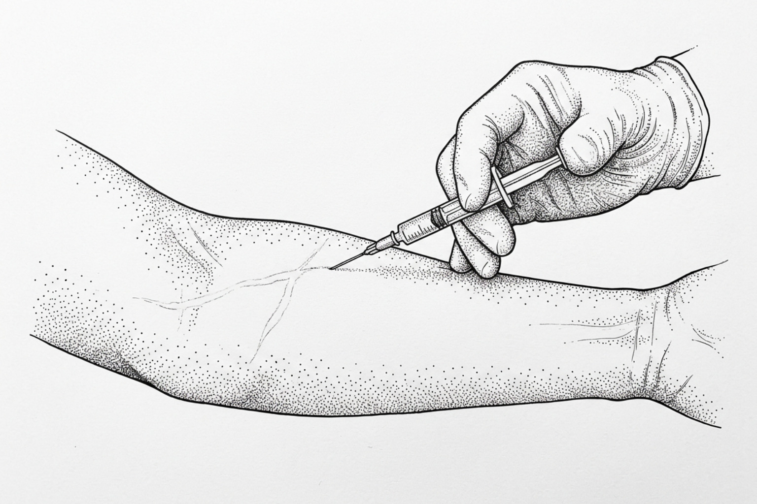
Preventing adverse reactions depends heavily on procedural accuracy, dosage selection, and proper post-treatment care.
Technique and dose choices that help
Refining injection technique helps ensure that the active medication remains confined entirely within the abnormal scar tissue matrix.
- Dose Fractionation and Concentration: Rather than delivering a single large bolus of high-concentration steroid (such as 40 mg/mL), clinicians often use lower concentrations (10 to 20 mg/mL) or titrate down to 2.5 to 10 mg/mL for smaller hypertrophic scars. Dosing protocols often deliver roughly 0.1 mL of suspension per 5 to 8 mm of linear scar tissue. Fractionating the total dose over multiple sessions spaced 4 to 6 weeks apart gives the tissue time to respond and lowers the risk of dermal thinning.
- Needle Gauge and Depth: Using fine-gauge needles (27-gauge to 30-gauge) allows for controlled tactile feedback. The clinician aims for the firm mid-dermis of the scar, stopping injection if loss of resistance suggests the needle has entered the softer subcutaneous fat layer.
- Saline Infiltration for Reversal: If persistent steroid-induced atrophy, depressions, or hypopigmentation develop, peer-reviewed clinical case studies demonstrate that serial intralesional injections of bacteriostatic 0.9% normal saline can help reverse these changes. The mechanical infiltration of saline helps resuspend the crystalline steroid aggregates. This allows local tissue macrophages to engulf and clear the drug depot, which can restore normal fibroblast function and epidermal volume over 2 to 6 months of treatment.
- Managing Telangiectasia and Deep Depressions: For telangiectasias that do not resolve on their own, pulsed dye lasers (585 or 595 nm) or intense pulsed light (IPL) can selectively target and coagulate dilated superficial vessels. In cases of severe, long-standing subcutaneous fat atrophy, corrective options include temporary hyaluronic acid soft-tissue fillers or autologous fat grafting.
Safer alternatives to consider
To minimize the high cumulative doses linked to monotherapy side effects, clinicians frequently combine corticosteroids with other modalities or evaluate alternative approaches.
| Scar Therapy Modality | Mechanism of Action | Common Local Side Effects | Risk of Systemic Complications | Scar Flattening Efficacy |
|---|---|---|---|---|
| Triamcinolone Acetonide (TAC) | Glucocorticoid receptor binding; collagenase activation; fibroblast suppression. | Skin atrophy (up to 63%), hypopigmentation, telangiectasia, pain. | Very Low to Moderate (depends on cumulative dose). | 70% – 80% response; 9% – 50% recurrence. |
| TAC + 5-Fluorouracil (5-FU) | Steroid anti-inflammatory action combined with a pyrimidine analog that halts DNA synthesis in fibroblasts. | Mild injection site pain, transient hyperpigmentation, superficial ulceration; lower atrophy rate than TAC alone. | Extremely Low (doses are well below systemic chemotherapy levels). | High (~92% volume reduction vs. 73% for TAC alone). |
| Intralesional Cryotherapy | Direct cellular freezing via cryoneedle; microvascular thrombosis and physical cell lysis. | Post-procedure blister formation, temporary edema, hypopigmentation (higher risk in darker skin phototypes). | None. | High (up to 75% local response; lower recurrence). |
| Intralesional Verapamil | Calcium channel blocker; increases procollagenase synthesis and alters cell shape. | Mild local burning, temporary erythema; significantly lower risk of atrophy or spider veins. | Very Low (minimal cardiovascular effects at scar doses). | Moderate to High (comparable long-term softening, slower onset). |
| Intralesional Vitamin D | Vitamin D receptor binding; modulation of fibroblast proliferation and collagen synthesis. | Injection site pain; significantly lower rates of skin atrophy and pigmentation change than TAC. | Very Low. | Lower than TAC. A 2026 meta-analysis of four randomized trials found TAC superior for scar severity and flattening. |
| Topical Silicone Gel / Sheets | Stratum corneum hydration; regulation of epidermal-dermal signaling and capillary activity. | Mild contact dermatitis, localized heat rash, superficial folliculitis. | None. | Mild to Moderate; ideal for early prevention and small scars. |
Evidence shows that combining low-dose triamcinolone with 5-fluorouracil provides higher scar reduction rates (around 92% compared to 73% for steroid monotherapy) while significantly reducing the incidence of dermal thinning and telangiectasia.
For patients seeking conservative, needle-free approaches, starting with a hypertrophic scar silicone gel or exploring non-invasive keloid removal pathways can help prevent and manage early raised scars without the side effects associated with injections.
Common questions
How long do these side effects last?
In many cases, steroid-induced skin thinning and pigment loss resolve on their own as the crystalline medication is gradually cleared from the tissue. This natural recovery typically takes 6 to 12 months, though some cases may take up to two years.
Because corticosteroids functionally inhibit melanocytes rather than destroying them, repigmentation usually occurs gradually as cell function returns. If white crystal deposits remain in the skin or the indentation does not improve after several months, serial normal saline infiltration performed by a dermatologist can help speed up drug clearance and tissue recovery.
Can repeated injections cause more scarring?
Yes. While the corticosteroid solution itself is designed to degrade collagen, the physical needle puncture causes mechanical micro-trauma.
If multiple injections are given too frequently into the exact same entry point using thick-gauge needles, this repetitive physical injury can stimulate localized wound healing and reactive fibrosis. In clinical practice, clinicians rotate injection puncture sites, use fine 30-gauge needles, and space appointments at least 4 to 6 weeks apart to prevent secondary needle-track scarring.
How can I avoid dents and visible veins?
The most reliable prevention strategy involves using conservative steroid concentrations (10 to 20 mg/mL for body keloids, and lower concentrations for facial or delicate skin), keeping the needle strictly within the firm mid-dermis of the scar, and avoiding over-injection into adjacent healthy skin or underlying fat.
Many dermatologists also use combination protocols—such as pairing low-dose triamcinolone with 5-fluorouracil or pulsed dye laser therapy—which helps flatten the scar while keeping the total steroid exposure below the threshold that triggers atrophy and telangiectasia.
What to take away
Intralesional corticosteroid injections remain a well-established, effective tool for flattening dense hypertrophic scars and relieving chronic itching and discomfort. However, because corticosteroids non-selectively suppress fibroblast activity and cellular turnover, localized side effects—including dermal thinning, skin depressions, hypopigmentation, and telangiectasia—occur in a notable percentage of patients. Systemic complications like adrenal suppression or Cushing's syndrome are rare, but risk levels rise with higher cumulative doses, frequent sessions, or treatment in pediatric populations.
Minimizing these risks relies on conservative drug concentrations, precise mid-dermal needle placement, adequate intervals between sessions, and the use of combination therapies when appropriate. If you are managing an elevated, painful, or changing scar and want personalized guidance on safe treatment options, you can complete a Scar Healing scar assessment to evaluate your scar and discuss the most appropriate path forward with a qualified specialist.
This content is for informational purposes only and does not constitute medical advice. Consult a qualified healthcare professional for diagnosis and treatment.
Works Cited
- British Association of Dermatologists. “Intralesional steroid therapy.” British Association of Dermatologists patient information, 2024.
- Gauglitz GG. “Management of keloids and hypertrophic scars: current and emerging options.” Clinical, Cosmetic and Investigational Dermatology, 2013.
- Jiang D, Liu Y, Li S, Wang X, You S. “Intralesional vitamin D versus triamcinolone acetonide for the treatment of keloids: a systematic review and meta-analysis of randomized controlled trials.” Frontiers in Medicine, 2026;13:1823636.
- Lee J, Kim J. “Minimal-invasive technologies for treatment of HTS and keloids: corticosteroids.” Textbook on Scar Management, Springer, 2020.
- Queen Victoria Hospital NHS Foundation Trust. “Corticosteroid injections in scars for adults and children.” QVH clinical leaflet 0670, 2022.
- Royal Free London NHS Foundation Trust. “Triamcinolone acetonide intradermal injection 10mg/ml for keloid and hypertrophic scars.” Royal Free London patient information, 2023.
- Sidhu G, Preuss CV. “Triamcinolone.” StatPearls, NCBI Bookshelf, 2026.
- Son D, Harijan A. “Overview of surgical scar prevention and management.” Journal of Korean Medical Science, 2014.
- Tullington JE, Gemma R. “Scar revision.” StatPearls, NCBI Bookshelf, 2026.