A Comprehensive Guide to Effective Keloid Scar Treatments
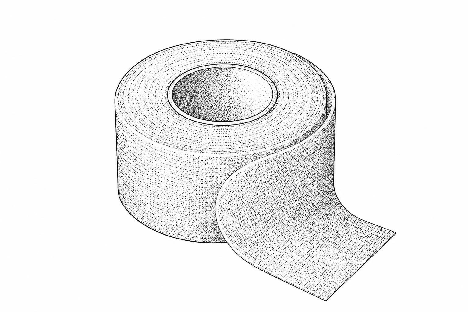
Keloids rarely go away on their own, and surgery without follow-up treatment often sees them return larger. Compare steroid injections, silicone sheeting, laser therapy, and surgery with radiation.
What the evidence says about keloid treatment
Treatment for keloid scars typically requires a multimodal approach — no single method works for everyone, and recurrence is common. Here is a quick overview of the main options ranked by evidence strength:
| Treatment | Evidence Level | Key Stat |
|---|---|---|
| Surgical excision + radiotherapy | Strong | Reduces recurrence to ~20% |
| Intralesional corticosteroids | Strong | 50–100% response rate |
| Silicone gel sheeting | Moderate | Up to 90% improvement reported |
| 5-Fluorouracil (5-FU) injections | Moderate | Effective in 45–96% of patients |
| Cryotherapy | Moderate | 30–75% success after two sessions |
| Pressure therapy | Low–Moderate | Requires 12–24 hrs/day for months |
| Onion extract (topical) | Weak | May ease itching; no proven removal |
| Surgery alone | Not recommended | 45–100% recurrence rate |
Keloids are among the most persistent and frustrating skin conditions in clinical dermatology. They are raised, firm scars that grow beyond the boundaries of the original wound — and unlike most scars, they do not fade or shrink on their own over time.
What makes them particularly difficult to manage is not just their physical nature. An estimated 100 million people worldwide acquire significant scars from surgery or trauma each year, with roughly 15% developing into excessive scarring. For those with darker skin tones, the risk is substantially higher — individuals with darker complexions are approximately seven times more likely to develop keloids than those with lighter skin.
Beyond the physical discomfort — which can include itching, tenderness, and restricted movement — keloids frequently carry a psychological toll. Visible scars in prominent locations can affect self-esteem, social confidence, and daily quality of life in ways that are often underestimated in clinical settings.
The good news is that evidence-based treatments do exist. The challenge lies in selecting the right combination for each individual case.
The Scar Healing Editorial Team has synthesized the current peer-reviewed clinical literature to walk through every major treatment pathway — from conservative at-home options to surgical and radiotherapy protocols — so readers can approach this topic with realistic, evidence-grounded expectations.
This content is for informational purposes only and does not constitute medical advice. Consult a qualified healthcare professional for diagnosis and treatment.
What keloids are and how they form
To understand why a treatment for keloid scars succeeds or fails, it is first necessary to examine the underlying biological dysfunction. Keloids represent a fibroproliferative disorder characterized by an abnormal wound healing response.
During normal wound healing, specialized dermal cells called fibroblasts synthesize collagen to rebuild the structural matrix of the skin. Once the wound is closed, a highly regulated balance of collagen synthesis and degradation restores tissue integrity. In keloid formation, this regulatory pathway fails. Fibroblasts remain in a prolonged, hypermetabolic state, continuously depositing excessive amounts of extracellular matrix proteins—primarily Type I and Type III collagen.
This cellular overactivity is often localized to anatomical zones subject to high mechanical skin tension, such as the anterior chest, shoulders, upper back, neck, and earlobes. However, systemic and genetic factors also drive this process. For example, conditions like Rubinstein-Taybi syndrome are genetically linked to a higher incidence of keloids.
Furthermore, individuals with darker skin phototypes (Fitzpatrick scales IV through VI) experience a significantly higher prevalence of keloid formation, with clinical estimates showing a 10% prevalence in African-American populations. For a deeper analysis of these biological mechanisms, refer to the complete guide to keloid causes.
Keloid or hypertrophic scar?
Differentiating between keloids and hypertrophic scars is a critical step in clinical dermatology, as the prognosis and treatment pathways differ substantially.
Hypertrophic scars generally appear within four weeks of a cutaneous injury, remain strictly confined within the original boundaries of the wound, and often undergo spontaneous regression within a year. Keloids, conversely, can develop months or even a year after the initial trauma. They actively invade surrounding healthy dermal tissue, show no tendency for spontaneous regression, and carry an exceptionally high risk of recurrence after intervention.
| Clinical Feature | Hypertrophic Scars | Keloids |
|---|---|---|
| Anatomical Boundaries | Remains within original wound borders | Extends beyond original wound borders into healthy skin |
| Onset Timeline | Typically within 4 weeks post-injury | Months to over a year post-injury |
| Spontaneous Regression | Often regresses or flattens over 12–18 months | Rarely regresses; may grow continuously |
| Anatomical Distribution | Any body site; common across joints | Predominantly chest, shoulders, earlobes, upper back |
| Recurrence After Surgery | Low recurrence rate | High recurrence rate (45% to 100% if untreated) |
According to the Merck Manual professional guidelines, a visual and physical examination is typically sufficient to distinguish between these two pathologies, though a skin biopsy may be utilized if the diagnosis remains uncertain.
Non-surgical keloid treatments
Conservative, non-invasive therapies are generally prioritized for early-stage scars, minor lesions, or as preventative measures immediately following wound closure. The primary therapeutic mechanisms of non-invasive options rest on occlusion and physical hydration of the stratum corneum.
When selecting conservative pathways, patients often explore various non-surgical keloid treatment options to avoid the risks associated with invasive procedures.
Silicone sheets and compression therapy
Silicone gel sheeting is widely accepted as a first-line, evidence-based non-surgical therapy. By creating an occlusive barrier, silicone sheets significantly reduce trransepidermal water loss (TEWL). This reduction in water loss restores hydration to the stratum corneum, which downregulates the signaling pathways that stimulate fibroblasts to produce excess collagen. Peer-reviewed clinical studies indicate that consistent application of silicone gel sheeting can yield up to a 90% improvement in the texture, pliability, and color of keloid scars.
Pressure therapy works alongside hydration to physically alter scar remodeling. Custom-fitted pressure garments or specialized compression clips (such as pressure earrings for earlobe keloids) exert continuous mechanical force on the scar tissue. This pressure creates localized hypoxia, which reduces cell viability, limits fibroblast proliferation, and accelerates collagen breakdown.
For clinical efficacy, pressure garments must exert a force of 24–30 mmHg and be worn continuously for 12 to 24 hours per day over a period of 4 to 6 months. To learn more about physical scar management, read about non-invasive keloid removal.
Creams and onion extract (Allium cepa) gels
Over-the-counter topical creams and gels frequently incorporate botanical active ingredients, most notably onion extract (Allium cepa). The primary active component in onion extract is quercetin, a bioflavonoid with known antihistaminic, anti-inflammatory, and antioxidant properties.
In vitro research demonstrates that quercetin can inhibit fibroblast proliferation and decrease collagen production. While onion extract can improve the overall cosmetic appearance of a scar and alleviate subjective symptoms such as pruritus (itching), clinical evidence does not support its use as a standalone method for removing established keloids.
According to a comprehensive Forum Dermatologicum review on non-surgical strategies, botanical topicals are best utilized as adjunctive therapies alongside more robust physical or pharmacological interventions.
Injections and in-office procedures
When conservative therapies fail to reduce the volume or symptoms of a keloid, invasive clinical treatments are required. Because keloid tissue exists in a cellular hypermetabolic state, targeting the rapidly proliferating fibroblasts with localized pharmacological agents can effectively arrest scar growth.
Steroid injections: the first-line treatment
Intralesional corticosteroid injection is the most widely utilized and clinically validated non-surgical treatment for keloid scars. The primary agent used is triamcinolone acetonide (TAC), typically administered at concentrations ranging from 10 to 40 mg/mL.
Corticosteroids work by suppressing localized inflammation, inhibiting fibroblast proliferation, and downregulating collagen synthesis. Additionally, they decrease the expression of endogenous collagenase inhibitors, promoting the natural enzymatic degradation of existing collagen fibers.
Clinical data shows that intralesional TAC injections achieve a response rate of 50% to 100%. However, the recurrence rate following steroid monotherapy ranges from 9% to 50% within five years.
To improve outcomes, clinicians often combine corticosteroids with antineoplastic agents like 5-fluorouracil (5-FU) in a 9:1 ratio (0.9 mL of 50 mg/mL 5-FU to 0.1 mL of 40 mg/mL TAC). This combination significantly enhances scar flattening while minimizing localized corticosteroid side effects such as dermal atrophy, telangiectasia (spider veins), and hypopigmentation.
A detailed review of keloid therapeutic management highlights that multimodal pharmacological regimens consistently outperform single-agent steroid therapies.
Laser treatment for keloids
Laser therapy offers a targeted approach to managing keloid vascularity and collagen structure. The most common modalities include:
- Pulsed-Dye Laser (PDL): Operating at a wavelength of 585 or 595 nm, the PDL specifically targets oxyhemoglobin within the microvasculature of the scar. This induces selective photothermolysis, destroying the blood vessels feeding the keloid, which starves the active tissue of oxygen and nutrients. PDL is highly effective at reducing scar erythema (redness) and alleviating pruritus.
- Fractional Ablative Lasers (CO2 and Er:YAG): These lasers create microscopic vertical channels in the scar tissue, a process known as fractional resurfacing, which stimulates healthier tissue remodeling.
- Laser-Assisted Drug Delivery (LADD): By utilizing a fractional CO2 laser to create microscopic channels immediately before applying a topical corticosteroid or 5-FU, clinicians can achieve uniform drug penetration throughout the deep dermis without the pain of direct needle injections.
To explore these light-based options, see the dedicated guide on laser therapy for keloids.
Surgery plus radiation
For large, debulking lesions or highly symptomatic keloids, surgical removal is often the most direct pathway to restore normal skin contours. However, performing surgery on a keloid patient carries a unique clinical risk: the surgical incision itself is a new cutaneous wound that can trigger an even larger keloid to grow back.
Why surgery alone often fails
Surgical excision performed as a standalone therapy (monotherapy) is highly discouraged in modern dermatology. Because the underlying fibroproliferative pathway remains active, excision alone carries an exceptionally high recurrence rate of 45% to 100%.
To minimize the risk of regrowth, surgeons must use meticulous techniques, such as intramarginal fusiform excision (leaving a tiny rim of the keloid border to avoid wounding healthy adjacent skin) and tension-free wound closure utilizing everted borders.
As detailed in a Frontiers systematic review on scar therapies, combining surgical excision with postoperative adjuvant therapies is essential to lower these high recurrence rates.
Radiation after surgery: timing and safety
Adjuvant postoperative radiation therapy is currently the most effective method for preventing keloid recurrence after surgical excision. Radiation works by inducing cellular apoptosis in rapidly dividing fibroblasts, which prevents the sudden surge of collagen synthesis that typically occurs during the early phases of wound healing.
The primary radiation modalities include:
- Superficial Radiation Therapy (SRT): Delivers low-energy X-rays that penetrate only the superficial layers of the skin, sparing deeper tissues.
- Electron Beam Therapy: Uses external high-energy electrons to deliver a precise dose to the surgical site.
- Brachytherapy: Involves placing a temporary radiation source (such as Iridium-192) directly into or adjacent to the surgical wound. Post-excisional brachytherapy has the lowest reported recurrence rate among all radiation modalities at approximately 15%.
For optimal clinical efficacy, the first fraction of radiation must be administered within a tight 24-to-48-hour window following surgical closure. According to the StatPearls clinical guide on radiation therapy, combining surgical excision with immediate postoperative radiotherapy reduces the keloid recurrence rate to approximately 20% overall, and to as low as 0% to 8.6% in highly controlled clinical trials.
While the risk of secondary radiation-induced malignancy is minimal in adults, radiation therapy is generally avoided or used with extreme caution in pediatric patients.
New treatments in development
As researchers uncover the precise cellular pathways of fibrogenesis, several novel molecular therapies are moving from clinical trials into practice:
- Transforming Growth Factor-Beta (TGF-β) Inhibitors: TGF-β1 and TGF-β2 are key cytokines that drive collagen synthesis, whereas TGF-β3 promotes scarless healing (similar to fetal skin). Recombinant human TGF-β3 (Avotermin) is under investigation to help prevent scarring.
- Calcium Channel Blockers: Intralesional verapamil blocks extracellular calcium influx, which downregulates interleukin-6 and collagen production while increasing collagenase activity.
- Immunomodulators: Topical imiquimod 5% cream, applied post-surgery, stimulates local interferon production, which increases collagen breakdown.
- Antineoplastic Repurposing: Intralesional bleomycin, a cytostatic antibiotic, is highly effective at flattening stubborn keloids, achieving success in up to 90% of patients with a low pooled recurrence rate of just 3%.
Botulinum toxin to ease skin tension
Botulinum Toxin Type A (commonly known as Botox) is emerging as a valuable tool in modern scar management. While traditionally used for cosmetic wrinkle reduction, its mechanism in keloid therapy is twofold.
First, by temporarily paralyzing the muscles surrounding a healing wound, Botulinum Toxin Type A significantly reduces the mechanical shearing tension on the wound edges. This tension reduction deprives local fibroblasts of the mechanical signals that trigger excessive collagen deposition.
Second, recent molecular studies demonstrate that Botulinum Toxin Type A directly inhibits fibroblast proliferation, downregulates profibrotic genes (such as TGF-β1), and reduces the expression of localized inflammatory mediators that cause pain and itching.
Diagnosing and preventing keloids
A formal clinical diagnosis is the first step toward managing a suspected keloid. Dermatologists typically perform a comprehensive physical examination and review the patient's medical history to evaluate the scar's growth pattern, texture, and symptoms.
If a lesion exhibits atypical features, or if there is any suspicion of a cellular malignancy, a skin biopsy is performed. A biopsy allows dermatopathologists to examine the tissue under a microscope to confirm the diagnosis and rule out other conditions.
Prevention if you're keloid-prone
For individuals with a personal or familial history of keloid formation, prevention is the most effective management strategy. The primary rule of prevention is to avoid any unnecessary cutaneous trauma, including elective cosmetic surgeries, body piercings, and tattoos.
When skin injuries or necessary surgical incisions do occur, high-risk individuals should initiate preventative protocols immediately after wound closure:
- Tension Reduction: Minimize physical activity that stretches the healing skin.
- Early Occlusion: Apply silicone gel patches or medical-grade silicone sheets daily for 12 to 24 hours.
- Topical Support: Use specialized formulations, such as a keloid scar prevention cream, to keep the healing tissue hydrated and calm.
Common questions about keloids
Do keloid scars go away on their own?
No. Unlike hypertrophic scars, which often undergo spontaneous regression and flatten over a period of 12 to 18 months, keloids are permanent tissue changes. They do not fade or regress on their own and typically require clinical intervention to shrink or flatten.
Is radiation therapy safe for treating keloids?
Yes, modern postoperative superficial radiation therapy is considered highly safe for adults. The radiation doses used are localized to the dermis, and the risk of secondary malignancy is minimal. However, it is generally avoided in pediatric patients due to the theoretical long-term risks to developing tissues.
Can keloids be permanently cured?
While "cure" is a difficult term in keloid management due to the high risk of recurrence, long-term remission is highly achievable. Utilizing multimodal therapies—such as combining surgical excision with postoperative radiation, or pairing steroid injections with pulsed-dye lasers—reduces recurrence rates to their lowest clinical levels, helping patients maintain flat, symptom-free skin.
Key takeaways
Successfully managing a keloid requires moving away from single-modality treatments. Because these lesions are driven by complex cellular pathways, a customized, multimodal approach tailored to the patient’s skin phototype, the scar's anatomical location, and its treatment history offers the highest chance of long-term success.
Whether utilizing conservative at-home therapies, localized pharmacological injections, or advanced post-surgical radiation, clinical evidence shows that combining treatments is key to preventing recurrence.
To find the right path for your skin, explore the clinical resources at keloid scar solutions, take a personalized scar assessment, or subscribe to our newsletter for the latest updates in evidence-based scar therapy.
Works Cited
- Kim SW. "Management of Keloid Scars: Noninvasive and Invasive Treatments." Archives of Plastic Surgery, vol. 48, no. 2, 2021, pp. 149–157.
- Ekstein SF, Wyles SP, Moran SL, Meves A. "Keloids: A Review of Therapeutic Management." International Journal of Dermatology, 2021.
- StatPearls. "Radiation Therapy in the Treatment of Keloids." StatPearls Publishing, NCBI Bookshelf.
- Merck Manual Professional Edition. "Keloids." Merck Manuals.
- Shen Y, Yang L, Feng D, et al. "Therapeutic Methods and Effect on Keloid and Hypertrophic Scars: A Systematic Review." Frontiers in Medicine, 2026.
- Zakrzewski B, Bednarski IA, Szala K, et al. "Non-surgical keloid management - review of established and emerging treatment strategies." Forum Dermatologicum, 2026.
This content is for informational purposes only and does not constitute medical advice. Consult a qualified healthcare professional for diagnosis and treatment.