Everything You Need to Know About Keloid Scar Solutions
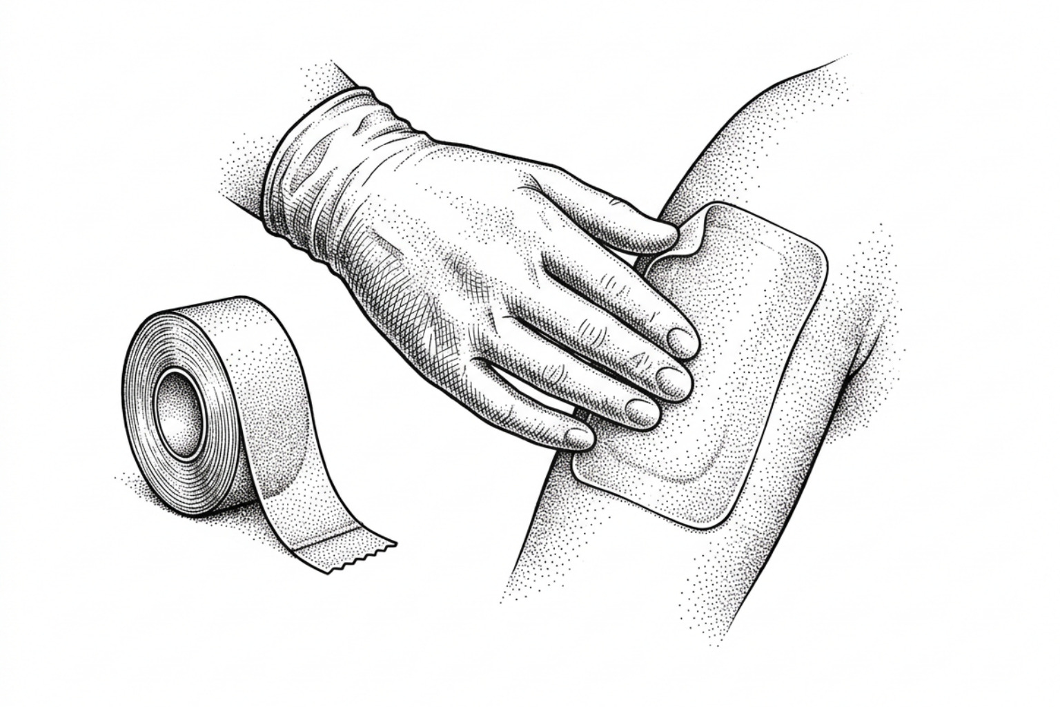
Keloids are stubborn and prone to recurrence. Here's how silicone, injections, lasers, surgery, and radiation compare, and how to choose the right one.
How keloid treatments compare
Keloid scar solutions range from first-line silicone therapy and corticosteroid injections to laser treatment, surgical excision, and emerging molecular therapies. No single treatment eliminates keloids permanently, but combination approaches consistently produce the best outcomes.
Quick reference — evidence-based keloid treatments by tier:
| Treatment Tier | Examples | Best For |
|---|---|---|
| Prevention / early management | Silicone gel or sheets, pressure garments | New wounds, post-surgical scars |
| First-line non-surgical | Intralesional corticosteroids (TAC), 5-FU | Established keloids of any size |
| Second-line / combination | Bleomycin, botulinum toxin A, laser therapy | Recalcitrant or symptomatic keloids |
| Procedural / surgical | Cryosurgery, excision + radiotherapy | Large, refractory, or pedunculated lesions |
Not in the table above: RNA interference and mesenchymal stem cell therapy are under investigation for keloids and are not standard care. Neither has human outcome data to grade, so this site describes them rather than rating them. Nothing in this category is available to patients today.
Keloids are among the most frustrating conditions in dermatology — not because they are dangerous, but because they are so difficult to resolve. Unlike a normal scar, which gradually flattens and fades, a keloid continues to grow beyond the original wound boundary, driven by persistent fibroblast activation and uncontrolled collagen overproduction. The result is a raised, firm, often itchy or painful lesion that can develop months or even years after the initial skin injury.
Keloids do not resolve on their own. According to clinical evidence reviewed across multiple prospective studies, recurrence rates after surgical removal alone reach 50–80% — and some sources cite figures approaching 100% without adjuvant therapy. This is not a condition where a single cream or one clinic visit delivers a lasting result.
What makes this topic genuinely complex is that keloid biology varies between individuals. Genetic predisposition, skin tone, the anatomical location of the scar, and the age of the lesion all influence how a keloid behaves — and which treatments are likely to work, or cause harm. People with Fitzpatrick skin types IV–VI (darker skin tones) face a disproportionate burden: they are more susceptible to keloid formation and also at higher risk of adverse effects such as post-inflammatory hyperpigmentation from certain treatments.
This guide brings together the current clinical evidence to explain what keloid scar solutions exist, how they work at a biological level, what the data actually shows about their effectiveness, and how clinicians and patients can think through a logical, stepwise approach to treatment.
This content is for informational purposes only and does not constitute medical advice. Consult a qualified healthcare professional for diagnosis and treatment.
Early treatment: silicone and pressure
Understanding how keloids form is essential to preventing them. Normal wound healing progresses through overlapping phases: inflammation, proliferation, and tissue remodeling. In keloid-prone individuals, this process becomes dysregulated.
During the proliferative and remodeling phases, fibroblasts produce an excessive amount of extracellular matrix, primarily type I and type III collagen. In a typical scar, collagen fibers eventually align parallel to the skin surface. In a keloid, the collagen fibers deposit in a disorganized, whorled, or haphazard pattern. For a detailed breakdown of this biological progression, see the guide on keloid scar formation stages.
While the exact genetic and environmental triggers remain under investigation, a thorough overview of known risk factors can be found in the keloid causes complete guide. Early intervention during the initial stages of wound closure is the most effective window for prevention.
Do silicone sheets work?
Topical silicone therapy is widely accepted as the gold standard for non-invasive scar management and prevention. Silicone gel or sheeting works primarily through occlusion and hydration rather than chemical absorption.
When applied to the stratum corneum (the outermost layer of the skin), silicone sheets or gels create a semi-occlusive barrier that reduces transepidermal water loss (TEWL). This localized hydration signals to the underlying epidermal cells that the barrier is intact, which in turn downregulates the production of pro-inflammatory cytokines. Consequently, fibroblast activity decreases, leading to a reduction in collagen synthesis.
According to a comprehensive review on Non-surgical keloid management review, silicone-based therapies are most effective when initiated immediately after wound epithelialization (closure) or suture removal. Clinical guidelines recommend:
- Daily wear time: 12 to 24 hours per day.
- Treatment duration: A minimum of 3 to 6 months.
- Format selection: Silicone gel sheets are highly effective for flat, stable areas, while self-drying silicone gels are preferred for mobile, highly visible, or contoured anatomical sites.
How pressure garments help
Pressure therapy relies on the principles of mechanotransduction—how physical forces influence cellular behavior. Applying continuous mechanical compression to a developing scar reduces local blood flow, creating a state of mild hypoxia. This localized oxygen deprivation limits the metabolic activity of fibroblasts, decreases collagen synthesis, and increases the activity of collagenase, an enzyme responsible for breaking down excess collagen.
Pressure garments and specialized appliances must be customized to provide a constant pressure of approximately 15 to 40 mmHg.
- Wear requirements: Patients must wear compression appliances for 12 to 24 hours per day (ideally up to 20 hours) for a duration of 4 to 6 months.
- Anatomical adaptations: For earlobe keloids—often caused by piercings—specialized pressure clip-on earrings are highly effective at preventing recurrence after surgical excision or during early-stage hypertrophic changes.
Injections for keloids
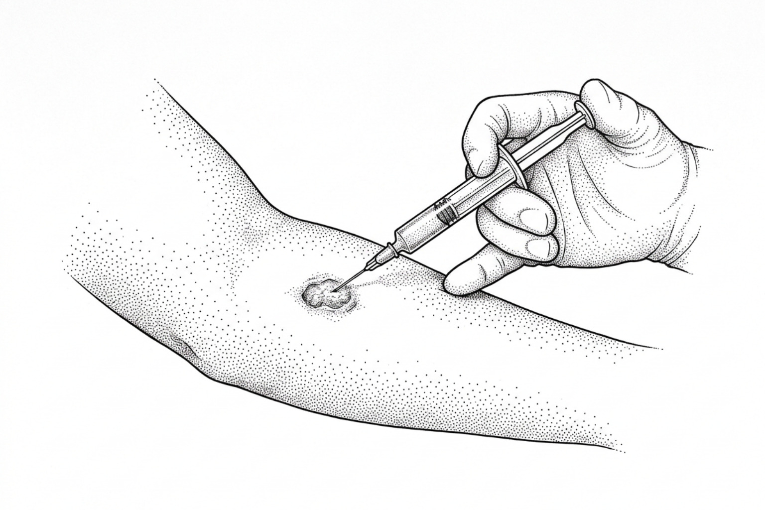
When a keloid is fully established, non-invasive topical therapies alone are rarely sufficient to flatten the lesion. In these cases, clinicians look to keloid treatment without surgery to target the overactive fibroblasts directly within the dermis. Intralesional pharmacotherapy remains the cornerstone of non-invasive keloid removal.
Steroid vs. chemotherapy injections
For decades, intralesional corticosteroid injections—most commonly triamcinolone acetonide (TAC)—have served as the first-line treatment for established keloids. TAC suppresses inflammation, inhibits fibroblast proliferation, and promotes the breakdown of collagen by downregulating collagenase inhibitors.
However, monotherapy with TAC has limitations. While 50% to 80% of keloids shrink after a series of monthly injections, a significant portion regrow within five years. Furthermore, repeated high-dose steroid injections can cause localized side effects, including skin atrophy, telangiectasia (visible blood vessels), and hypopigmentation.
To improve efficacy and minimize side effects, clinicians frequently combine or substitute corticosteroids with chemotherapeutic agents:
- 5-Fluorouracil (5-FU): This pyrimidine antagonist inhibits DNA synthesis, slowing down the rapid proliferation of keloid fibroblasts. Clinical evidence detailed in the Layered Management of Hypertrophic Scars and Keloids review demonstrates that combining TAC with 5-FU provides faster flattening, superior symptom relief, and better long-term durability than TAC alone, while reducing the risk of steroid-induced skin atrophy.
- Bleomycin: An antitumor antibiotic that directly inhibits collagen synthesis and induces fibroblast apoptosis (programmed cell death). Clinical trials show that intralesional bleomycin is highly effective, achieving a significant flattening rate of up to 90% in clinical trials, with an exceptionally low pooled recurrence rate of approximately 3%. The primary side effects include localized pain during injection and transient hyperpigmentation.
Botulinum toxin (Botox) injections
Botulinum Toxin A (BoNTA), commonly known for its cosmetic applications, has emerged as a valuable adjunctive treatment for keloids. The therapeutic mechanism is twofold:
- Tension Reduction: By paralyzing the localized skeletal muscles surrounding a wound or scar, BoNTA reduces the mechanical tensile force pulling on the healing tissue. Low-tension environments naturally synthesize less collagen.
- Cellular Downregulation: In-vitro studies suggest that BoNTA directly downregulates the expression of Transforming Growth Factor-beta 1 (TGF-β1), a key cytokine responsible for driving fibroblast hyperactivity.
According to the Evidence-based systematic review of recent advances, alternative intralesional agents such as verapamil (a calcium channel blocker) and hyaluronidase are also under investigation. Verapamil stimulates host collagenase production and inhibits extracellular matrix synthesis, presenting a steroid-free option with a lower risk of pigmentary changes.
Laser treatments for keloids
Energy-based devices offer precise options for addressing the vascularity, height, and texture of keloid scars. By targeting specific chromophores (such as hemoglobin in blood vessels), lasers can selectively destroy the microvasculature supplying nutrients to the overactive scar tissue. To understand the different laser technologies available, consult the comprehensive resource on laser for keloid scars.
Pulsed-Dye Laser (PDL) and Nd:YAG Systems
The Pulsed-Dye Laser (PDL), operating at a wavelength of 585 nm or 595 nm, is highly absorbed by oxyhemoglobin. This targeting results in photothermal destruction of the scar's microvasculature, leading to localized tissue hypoxia and a subsequent decrease in fibroblast activity. PDL is highly effective at reducing scar erythema (redness) and relieving associated pruritus (itching).
The 1064 nm Nd:YAG laser penetrates deeper into the dermis than the PDL, making it suitable for thicker, more substantial keloids. It also targets microvasculature, reducing cellular activity deep within the lesion. Both laser therapies are typically administered over multiple sessions spaced 4 to 8 weeks apart. They are rarely used as monotherapies for thick keloids; instead, they are combined with intralesional steroid injections to maximize tissue softening and flattening.
Fractional CO2 and Laser-Assisted Drug Delivery (LADD)
Ablative fractional lasers, such as the fractional carbon dioxide (CO2) laser, create thousands of microscopic, vertical thermal treatment zones (microchannels) in the skin while leaving the surrounding tissue intact. This process initiates a rapid wound-healing response that replaces disorganized keloidal collagen with organized, healthy collagen fibers. For those managing acne-induced lesions, a detailed clinical perspective is available in the microneedling acne keloid scars complete guide.
Beyond physical remodeling, fractional lasers are increasingly used for Laser-Assisted Drug Delivery (LADD). The microchannels created by the laser act as physical pathways, allowing topically applied medications (such as TAC or 5-FU) to penetrate deeply and uniformly into the dermis. As highlighted in the PMC Article on Keloid Management, LADD significantly improves the bioavailability of these therapeutics while reducing the intense localized pain and needle anxiety associated with traditional intralesional injections.
Surgery and radiation for keloids
Surgical removal of a keloid is a double-edged sword. Because surgical excision involves cutting into the skin, it triggers a new inflammatory cascade. Without immediate preventive measures, the body's abnormal wound-healing response is re-activated, resulting in a recurrence rate of 50% to 80%—and in some anatomical areas, nearly 100%. When surgical removal is necessary, such as in cases of large, pedunculated lesions or for abdomen keloid scar reduction, strict adherence to adjuvant (secondary) therapies is mandatory.
Radiation after surgery
To prevent the rapid recurrence of keloid tissue post-surgery, clinicians often utilize localized radiation therapy. Radiation targets and destroys rapidly dividing fibroblasts at the wound margin, preventing the overproduction of collagen during the early phases of healing.
- Superficial Radiation Therapy (SRT): Delivers precise, low-energy X-rays that penetrate only a few millimeters into the skin, minimizing exposure to deeper tissues.
- Strontium-90 (Sr-90) Beta-Brachytherapy: A highly localized form of radiation therapy that utilizes a radioactive applicator placed directly on the wound site. Because beta particles have a very short penetration depth (75% of Sr-90 radiation is absorbed within the first 2 mm of tissue), it is highly effective for thin, superficial lesions. Clinical data indicates that utilizing Sr-90 post-excision can halve the recurrence rate of stubborn keloids.
- The Critical Window: For radiotherapy to be successful, the first fraction of radiation must be administered within 24 hours of the surgical closure.
- Safety and Risks: While modern, highly targeted radiotherapy protocols are safe, clinicians must carefully weigh the treatment against potential long-term risks, including localized skin peeling, permanent pigmentary changes, and a theoretical, albeit very low, long-term oncogenic (cancer) risk.
Freezing and combination treatments
Cryosurgery utilizes liquid nitrogen to freeze and destroy cellular structures within the keloid. This freezing causes vascular thrombosis (clotting) and localized cell death, leading to the gradual sloughing off of the scar tissue.
- Contact vs. Intralesional Cryotherapy: Traditional contact or spray cryotherapy freezes the scar from the outside in, which can cause significant damage to the overlying epidermis. Intralesional cryotherapy utilizes a specialized needle probe inserted directly into the core of the keloid, freezing the lesion from the inside out. This preserves the surface melanocytes (pigment cells) and reduces healing times.
- Combination Protocols: Cryotherapy is frequently combined with intralesional TAC injections. Freezing the dense collagen matrix of a keloid makes the tissue softer and easier to inject, allowing for a more uniform distribution of the corticosteroid.
Choosing treatment for your skin type
No two keloids are identical, and a treatment plan that works for a small earlobe lesion may fail or cause severe complications if applied to a large chest keloid. Effective management requires a personalized approach that accounts for the patient's skin type, age, and the physical characteristics of the scar itself. To explore case studies and articles organized by specific scar profiles, browse the tag keloid scars resource library.
Treating keloids on darker skin
Patients with Fitzpatrick skin types IV through VI have a higher genetic susceptibility to keloids and are uniquely vulnerable to treatment-induced complications.
- Pigmentary Risks: Aggressive cryotherapy, high-dose corticosteroid injections, and certain laser wavelengths can destroy melanocytes, leaving behind permanent white patches (hypopigmentation) or dark, post-inflammatory hyperpigmentation (PIH) spots.
- Steroid-Sparing Strategies: For patients with darker skin tones, clinicians often prioritize steroid-sparing agents like 5-FU, verapamil, or low-dose combination therapies to avoid localized skin bleaching and dermal atrophy.
- Anatomical Considerations:
- Earlobe Keloids: Respond exceptionally well to surgical excision followed by pressure earrings and localized silicone gel.
- Chest and Joint Keloids: High-tension areas are highly prone to stretching and recurrence. These sites require robust tension-relieving surgical closures, long-term silicone sheets, and early adjuvant intralesional therapies.
- Age Factors: Keloids are most common in individuals aged 10 to 30. Pediatric patients require careful consideration, often starting with non-invasive silicone and pressure therapies before escalating to injections or energy-based devices.
New research and treatment plans
As our understanding of the molecular pathways driving keloid pathogenesis improves, researchers are developing targeted therapies designed to arrest keloid growth at the genetic and cellular levels.
Gene and stem cell therapies
- RNA Interference (siRNA): This technology utilizes small interfering RNA molecules to silence specific genes responsible for collagen production and tissue remodeling. Preclinical studies are targeting genes such as TIMP-1 (tissue inhibitor of metalloproteinases-1), Runx2, and circCOL5A1. Silencing these targets helps restore the balance of collagen degradation and synthesis within keloid fibroblasts.
- Mesenchymal Stem Cells (MSCs): Adipose-derived stem cells (ADSCs) are being studied for their immunomodulatory properties. In laboratory models, MSC therapies have shown the ability to downregulate the pro-fibrotic TGF-β1/Smad signaling pathway, reducing the expression of collagen types I and III, alpha-smooth muscle actin (α-SMA), and fibronectin.
Shock-wave therapy
- Extracorporeal Shock-Wave Therapy (ESWT): This non-invasive modality uses acoustic waves to stimulate mechanosensitive ion channels in the skin. ESWT has been shown to modulate the inflammatory phase of healing, downregulate pro-fibrotic cytokines, and promote healthier, more organized collagen remodeling without the need for steroid injections.
- Mechanotherapy in Surgery: Advanced surgical closure techniques, such as deep tension-relieving sutures and progressive tension sutures, are designed to offload mechanical forces from the healing wound edges, directly preventing the mechanotransduction cascade that triggers keloid recurrence.
A step-by-step treatment plan
To manage keloids safely and effectively, clinicians utilize a structured, stepwise approach, escalating from low-risk preventive measures to aggressive combination therapies for refractory (resistant) cases.
| Step | Phase / Scar Status | Primary Therapeutic Strategy | Expected Timeline |
|---|---|---|---|
| Step 1 | Prevention (post-injury/surgery) | Topical silicone gel/sheets + pressure garments | 12–24 hours/day for 3–6 months |
| Step 2 | Immature / Mild Keloid | Intralesional TAC (10-20 mg/mL) or TAC + 5-FU | Monthly injections for up to 6 months |
| Step 3 | Moderate / Symptomatic | Pulsed-Dye Laser (erythema) or LADD with TAC/5-FU | Sessions every 4–8 weeks |
| Step 4 | Large / Pedunculated | Surgical Excision + Adjuvant Radiotherapy (SRT/Sr-90) | Radiotherapy within 24 hours post-op |
| Step 5 | Refractory / Recurrent | Intralesional Bleomycin, Cryosurgery + TAC, or clinical trials | Case-by-case individualization |
Frequently asked questions
Why do keloid scars have such high recurrence rates after surgical removal?
Surgical excision removes the physical mass of the keloid, but it also creates a new wound. In individuals with a genetic predisposition to keloids, this new wound triggers the same abnormal, hyper-inflammatory healing response. Without immediate adjuvant therapies—such as radiation, pressure therapy, or steroid injections—to suppress the newly activated fibroblasts, the body will synthesize collagen at an accelerated rate, causing the keloid to return, often larger than the original lesion.
How does skin tone affect the safety and choice of keloid treatments?
Melanocytes (the cells responsible for skin pigment) are highly sensitive to temperature extremes, inflammation, and chemical trauma. Treatments like cryosurgery and high-dose corticosteroid injections can damage these cells, leading to permanent hypopigmentation (white spots) or post-inflammatory hyperpigmentation (dark spots) in patients with Fitzpatrick skin types IV–VI. To minimize these risks, clinicians utilize conservative treatment settings, test spots, and steroid-sparing agents like 5-FU or verapamil.
Can natural remedies like onion extract successfully flatten a keloid?
Onion extract contains compounds like quercetin and kaempferol, which possess natural anti-inflammatory and anti-proliferative properties. Some clinical studies suggest that topical onion extract gel can improve the overall appearance, redness, and associated itch or pain of hypertrophic scars and mild keloids. However, there is no strong clinical evidence demonstrating that onion extract alone can successfully flatten an established, thick keloid scar. It is best used as a mild, complementary home remedy alongside primary medical treatments like silicone therapy.
Key takeaways
Managing keloid scars requires a long-term, multimodal approach. Because keloids are characterized by chronic inflammation and a high propensity for recurrence, relying on a single treatment method is rarely successful. By combining early preventive measures like silicone occlusion with targeted medical interventions—such as intralesional pharmacotherapy, advanced laser systems, and selective radiotherapy—patients and clinicians can successfully control scar volume and alleviate symptoms.
To take the first step toward finding an individualized management plan, utilize the independent Scar Assessment Tool to evaluate your scar profile, or explore the science of tissue regeneration at Scar Healing.
Works Cited
- Zakrzewski B, Bednarski IA, Szala K, et al. "Non-surgical keloid management - review of established and emerging treatment strategies." Forum Dermatologicum, 2026.
- Abbadi OS, et al. "Layered Management of Hypertrophic Scars and Keloids: From Silicone and Intralesional Triamcinolone Acetonide Plus 5-Fluorouracil to Adjuvant Strontium-90." Cureus, 2026.
- Walsh LA, Wu E, Pontes D, et al. "Keloid treatments: an evidence-based systematic review of recent advances." Systematic Reviews, 2023;12(1):42.
- Murakami T, Shigeki S. "Pharmacotherapy for Keloids and Hypertrophic Scars." International Journal of Molecular Sciences, 2024;25(9):4674.
This content is for informational purposes only and does not constitute medical advice. Consult a qualified healthcare professional for diagnosis and treatment.