Intralesional corticosteroid for keloids: what the evidence shows.
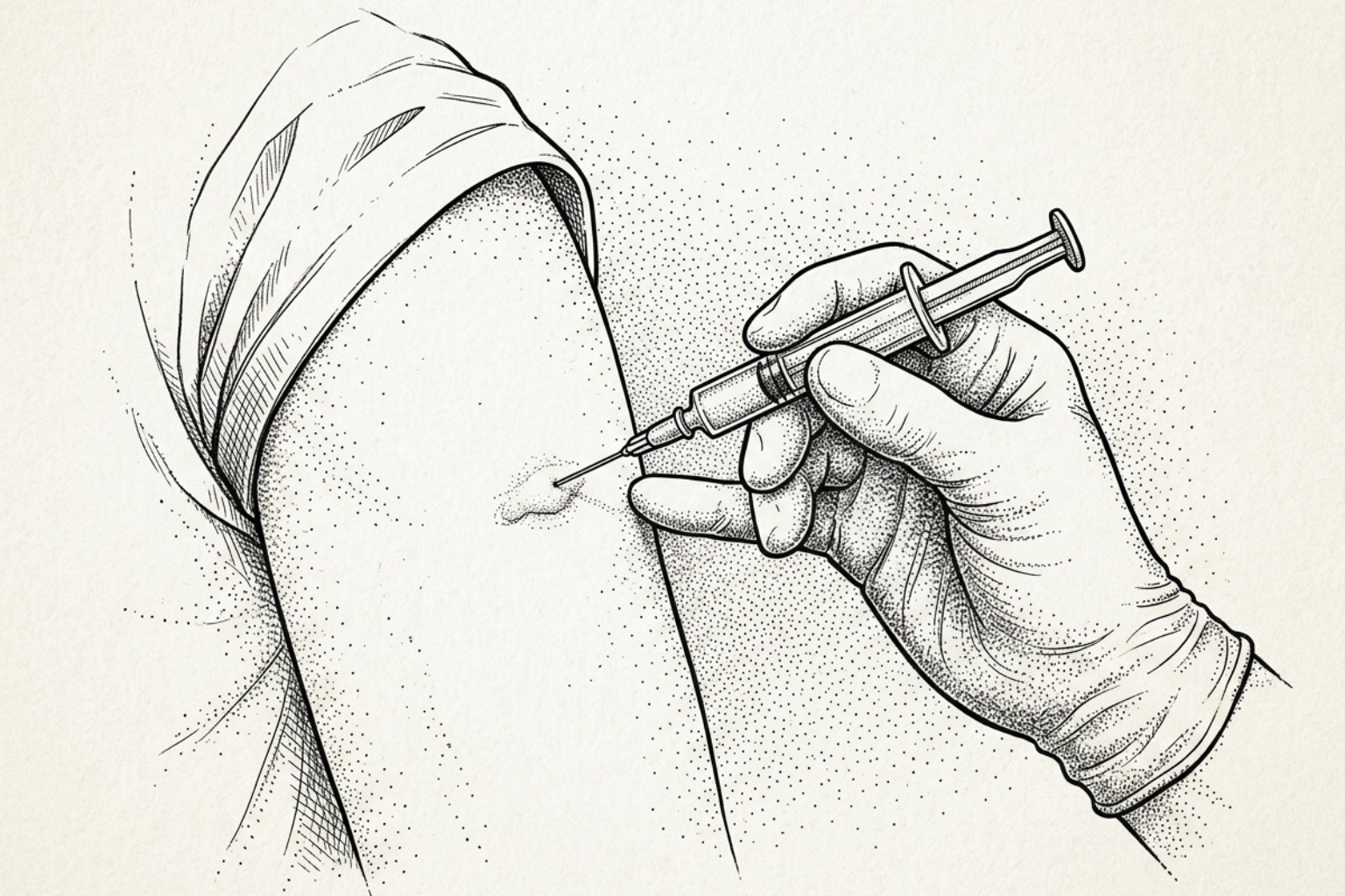
Intralesional triamcinolone is the first-line keloid treatment worldwide, yet response rates and recurrence vary widely. Here is what the dosing evidence and international consensus actually say.
Why steroid injections are used for keloids
Intralesional corticosteroid for keloids means injecting a steroid medicine directly into the raised scar. Evidence suggests that triamcinolone acetonide is the usual first-line option because it can soften and flatten keloid tissue while easing itch and pain. Treatment is normally given in a series of carefully placed injections, often about 3 to 4 weeks apart, by a qualified clinician.
Keloids are firm scars that grow beyond the original wound. They form when fibroblasts, the cells involved in skin repair, keep producing excess collagen and extracellular matrix. Research indicates that injected corticosteroids can reduce this activity, limit inflammation, and decrease the scar's blood supply. The goal is improvement, not a guaranteed cure: published outcomes vary widely, and keloids can return after treatment.
Triamcinolone acetonide was used in 37 of 38 randomized trials reviewed on intralesional corticosteroid treatment for keloids. Clinical research reports scar flattening in roughly 50% to 100% of cases, but recurrence estimates range from 9% to 50%, partly because studies use different doses, injection methods, and follow-up periods.
The sections ahead explain what the evidence says about dosing, injection technique, pain control, side effects, combination treatment, and realistic long-term monitoring.
This guide was prepared by The Scar Healing Editorial Team.
How the injection works, and how well
Understanding how abnormal scars form requires a close look at the cellular machinery driving tissue repair. In healthy wound healing, inflammation subsides once re-epithelialization is complete, allowing fibroblasts to balance collagen production with matrix remodeling. In keloids, this regulatory switch fails. Fibroblasts remain chronically active, synthesizing excessive type I and type III collagen fibers that aggregate into dense, hyalinized bundles extending beyond original wound borders.
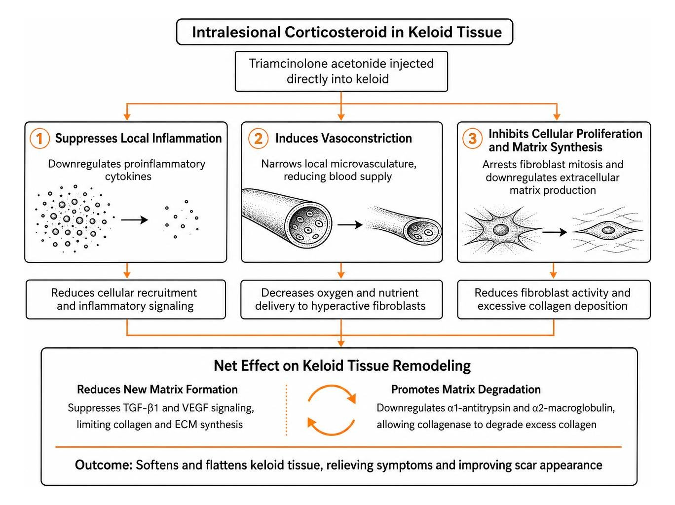
When clinicians administer an intralesional steroid, they deposit the medication directly into this hyperactive cellular environment. A comprehensive scoping review on intralesional corticosteroid injection methods demonstrates that localized steroid delivery alters aberrant scar biology through three principal actions:
- Suppression of local inflammation: Corticosteroids downregulate proinflammatory cytokines that perpetuate cellular recruitment.
- Vasoconstriction: Injected glucocorticoids induce local microvascular narrowing, reducing oxygen and nutrient delivery to hypermetabolic fibroblasts.
- Inhibition of cellular proliferation: Glucocorticoids arrest fibroblast mitosis and downregulate extracellular matrix synthesis.
What triamcinolone does to scar tissue
Triamcinolone acetonide (TAC) operates by diffusing across cell membranes and binding to intracellular glucocorticoid receptors. Once activated, these receptor complexes translocate to the nucleus, where they inhibit transcription factors such as nuclear factor-kappa B (NF-κB) and activator protein 1 (AP-1). This cellular cascade suppresses transformative signaling molecules, notably transforming growth factor-beta 1 (TGF-β1) and vascular endothelial growth factor (VEGF).
Beyond stopping the production of new matrix components, triamcinolone helps the body clear existing scar tissue. The drug downregulates the natural collagenase inhibitors alpha-1-antitrypsin and alpha-2-macroglobulin. By neutralizing these inhibitors, endogenous dermal collagenase can freely degrade excess collagen bundles. This dual mechanism—reducing new matrix deposition while facilitating the breakdown of existing fibrous tissue—helps explain why intralesional injections remain central to addressing underlying keloid causes.
How often it works
Published clinical response rates for intralesional triamcinolone acetonide monotherapy vary across clinical trials, with scar flattening generally documented between 50% and 100%. However, a patient's therapeutic outcome depends heavily on scar chronicity and mechanical properties.
In a comparative clinical study on intralesional steroids versus cryotherapy, researchers observed distinct response profiles based on how long the keloid had been present:
- Early lesions (duration ≤1 year): Achieved a 100% complete flattening rate after up to five sessions spaced 3 weeks apart.
- Intermediate lesions (duration >1 to ≤4 years): Demonstrated a 94.1% complete flattening rate, outperforming cryotherapy monotherapy (62.5%, p = 0.028).
- Longstanding lesions (duration ≥5 years): Displayed marked resistance to monotherapy, achieving complete flattening in only 15.4% of cases.
Standardized clinical evaluations using the Vancouver Scar Scale (VSS) and Patient and Observer Scar Assessment Scale (POSAS) show that symptomatic relief often precedes visible tissue flattening. Patients frequently report significant reductions in pruritus and pain within the first two treatment cycles as acute inflammatory mediator levels drop.
Dosing and how the injection is given
Achieving reliable outcomes with intralesional corticosteroids requires precise execution. Unlike systemic medications dosed strictly by body weight, intralesional protocols demand adjustments based on tissue density, anatomical site, and scar surface area. Implementing standardized dermatology scar management protocols helps minimize both under-treatment and operator-dependent complications.
Standard doses and concentrations
Triamcinolone acetonide concentrations used in clinical practice range from 10 mg/mL to 40 mg/mL. The international e-Delphi consensus from the KECORT Study: An International e-Delphi Study on the Treatment of KEloids Using Intralesional CORTicosteroids in Clinical Practice established that 40 mg/mL represents the standard first-line concentration for firm, elevated keloids on the trunk and extremities. Lower concentrations (typically 2.5 to 10 mg/mL) are preferred for delicate areas like the face or earlobes to reduce the risk of dermal thinning.
Published trials reflect significant dosing variability, with administered doses ranging from 1 mg to 20 mg of TAC per cm² of keloid tissue. However, to prevent systemic complications, clinical guidelines recommend establishing a firm monthly ceiling.
| Parameter | Clinical Consensus / Standard Range | Special Considerations |
|---|---|---|
| TAC Concentration | 10 to 40 mg/mL | 40 mg/mL for trunk/limbs; 2.5–10 mg/mL for facial lesions |
| Injection Volume per Site | 0.1 to 0.2 mL per cm² | Titrate carefully until blanching occurs |
| Maximum Dose per Session | 40 mg to 80 mg total cumulative dose | Do not exceed 80 mg/month to avoid adrenal suppression |
| Session Frequency | Every 3 to 4 weeks | 50% of RCT protocols use 4-week intervals; 29% use 3-week intervals |
| Course Duration | Median of 4 to 6 sessions | Discontinue once tissue reaches level skin margins |
Needle, depth and when to stop
Injecting into dense, fibrous scar tissue requires substantial mechanical force. Standard friction-fit (slip-tip) syringes carry a risk of needle dislodgement under high injection pressures. Clinical consensus recommends using 1 mL polycarbonate Luer-lock syringes, which secure the needle hub and allow fine volumetric control.
- Needle selection: Fine-gauge needles ranging from 25G to 30G minimize puncture trauma while generating adequate intralesional hydraulic pressure.
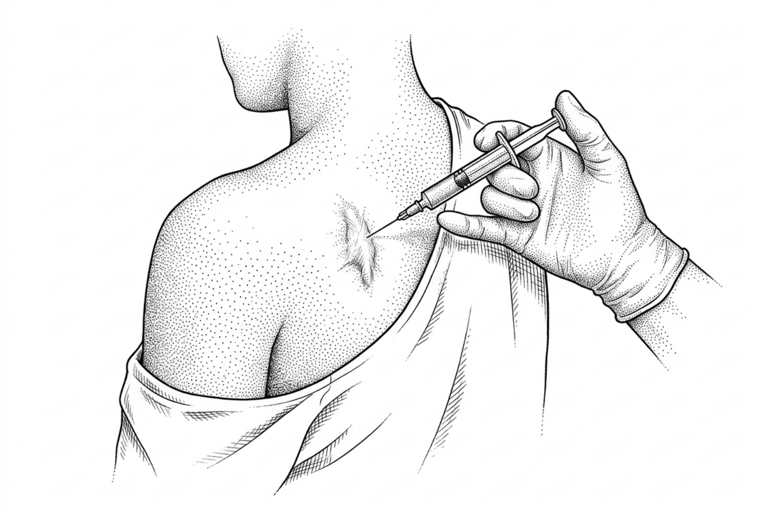
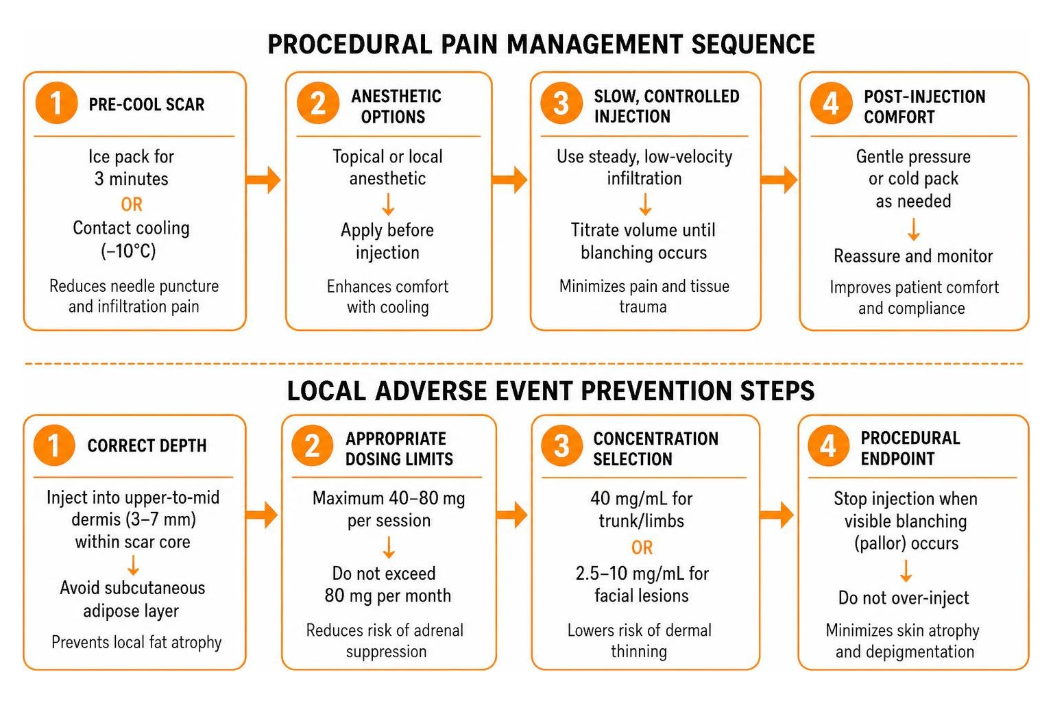
- Injection depth: The needle must target the dense, indurated core of the scar within the upper-to-mid dermis (typically 3 to 7 mm deep, depending on lesion elevation). Clinicians must avoid depositing medication into the subcutaneous adipose layer beneath the scar, as misdirected steroid crystals induce localized fat atrophy.
- Puncture distribution: For broad keloidal plaques, clinicians use a multi-puncture technique, spacing injection points roughly 1 cm apart across the surface.
- Procedural endpoint: Infiltration should stop immediately once visible tissue blanching (pallor) is observed across the treated segment. Blanching indicates that the crystalline suspension has thoroughly dispersed through the dense collagen matrix.
Pain and side effects
Intralesional injections into firm scars can cause notable discomfort. Pain levels correlate with scar density, injection velocity, and anatomical keloid body location risks, with presternal, palmar, and plantar locations presenting heightened sensitivity.
How to make it hurt less
Controlling procedural discomfort improves patient compliance and allows clinicians to perform deliberate, controlled injections without rushing.
- Skin pre-cooling: Research indicates that pre-cooling the scar surface with an ice pack for 3 minutes or applying a contact cooling device (−10°C) reduces needle puncture and infiltration pain more effectively than topical anesthetic creams alone. Cold therapy temporarily slows peripheral nerve conduction.
- Topical anesthetics: Eutectic mixtures of local anesthetics (e.g., 2.5% lidocaine / 2.5% prilocaine cream) applied under occlusion for 60 minutes prior to injection penetrate up to 5 mm into intact skin, diminishing superficial puncture pain.
- Sub-lesional local anesthetic blocks: Infiltrating 1% or 2% plain lidocaine into the looser subcutaneous tissue beneath the scar creates a regional field block while elevating the dense keloid, making subsequent intralesional needle insertion easier.
- Injection speed control: Injecting the suspension slowly (approximately 0.1 mL per 10 to 15 seconds) prevents sudden mechanical distension of rigid fibrous tissue.
- Admixture considerations: Although mixing lidocaine directly with triamcinolone in the same syringe remains common in outpatient practice, expert consensus panels (such as the KECORT study) advise against it, noting that direct admixtures dilute steroid concentration without eliminating initial needle puncture pain.
Avoiding thinning, veins and pale patches
Local adverse events occur frequently with intralesional corticosteroid therapy. Published reviews indicate that localized skin atrophy occurs in 5% to 80% of treated patients, while telangiectasias (prominent microvessels) appear in up to 80% of cases.
Hypopigmentation represents another major consideration, with clinical trials documenting pigmentary alterations in 12.5% to 75% of patients. Corticosteroids suppress melanocyte metabolism and melanin synthesis, an effect that is more pronounced in deeply pigmented skin (Fitzpatrick phototypes IV–VI).
According to the textbook on scar management and minimally invasive technologies, clinicians can limit these adverse effects through several practical safeguards:
- Strict intralesional confinement: Ensure all steroid deposition occurs within the scar margins, avoiding spillover into adjacent healthy dermis or subcutaneous fat.
- Lower baseline concentrations: Initiate therapy at lower concentrations (10 to 20 mg/mL) and increase to 40 mg/mL only if the scar demonstrates clinical resistance.
- Timely treatment cessation: Stop injections as soon as the keloid flattens to the level of surrounding skin; continued treatment past this point leads to epidermal depression.
- Managing secondary changes: If persistent telangiectasias develop after scar resolution, vascular lasers such as the 595 nm pulsed dye laser (PDL) can selectively photocoagulate superficial dilated vessels.
Combining steroids with other treatments
While intralesional corticosteroid monotherapy remains standard, recalcitrant or bulky scars often respond better to multimodal strategies. Exploring non-surgical keloid treatment options that combine complementary mechanisms helps clinicians achieve greater scar reduction while using lower cumulative steroid doses.
Adding 5-fluorouracil
Combining triamcinolone acetonide with the pyrimidine analog 5-fluorouracil (5-FU) is one of the most extensively studied combination approaches. As detailed in a scientific review on intralesional injection modalities for keloids, 5-FU blocks thymidylate synthase, arresting DNA synthesis and driving fibroblast apoptosis.
A standard formulation combines nine parts 5-FU (50 mg/mL) with one part triamcinolone acetonide (40 mg/mL), yielding a mixture containing 45 mg/mL of 5-FU and 4 mg/mL of TAC. This pairing offers distinct clinical advantages:
- Enhanced efficacy: Randomized trials report average scar volume reductions reaching 92% with the combination, compared to 73% for steroid monotherapy.
- Mutual mitigation of side effects: The antimetabolite action of 5-FU accelerates tissue remodeling, while the low-dose corticosteroid suppresses 5-FU-induced tissue inflammation and ulceration. Simultaneously, reducing the total steroid dose lowers the incidence of dermal atrophy and telangiectasia.
Cryotherapy and Laser-Assisted Drug Delivery
Physical modalities can alter the structural density of keloids, making drug delivery more efficient.
- Cryotherapy pre-treatment: Applying liquid nitrogen (open spray or contact probe for 10 to 30 seconds across 2 to 3 freeze-thaw cycles) induces intracellular ice formation and localized interstitial edema. Once the scar thaws, the softened tissue allows easier needle penetration and uniform steroid dispersal. Clinical studies show combining cryotherapy with intralesional triamcinolone yields response rates up to 84%.
- Laser-assisted drug delivery: Utilizing laser treatments for keloid scars, such as ablative fractional CO2 lasers, creates microscopic vertical ablation channels across the stratum corneum. Applying topical triamcinolone over these channels allows deep dermal diffusion without needle puncture pain.
- Radioporation: In an open-label randomised controlled trial on radioporation-assisted steroid delivery, radiofrequency-created micro-ports combined with topical triamcinolone produced a 68% scar volume reduction at 24 weeks with significantly less dermal atrophy (16%) than standard needle injection (29%, p < 0.05).
- Silicone elastomer sheeting: Placing medical-grade silicone gel sheets over the treated scar for 12 to 24 hours daily between injection sessions enhances stratum corneum hydration, downregulates growth factors, and protects against friction.
Intralesional vitamin D
Vitamin D has been studied as a lower-risk substitute for triamcinolone rather than a partner to it. A 2026 systematic review and meta-analysis of four randomized trials found triamcinolone more effective for both scar severity and lesion flattening, while vitamin D produced significantly less pigmentation change and significantly less skin atrophy. The trade is efficacy against side effects, which makes vitamin D worth raising for scars on visible areas, or if earlier steroid injections have already left thinning or pale patches.
Follow-up and the risk of return
Because the cellular drivers of abnormal tissue growth can persist long after clinical flattening, managing a keloid requires ongoing observation across the different stages of keloid scar formation.
Who should not have this treatment
Although intralesional injections limit systemic drug distribution, triamcinolone crystals gradually solubilize and enter peripheral circulation. Clinicians must observe key medical precautions before and during treatment:
- Active infection: Never inject into or adjacent to active bacterial, viral (e.g., herpes simplex), or fungal skin lesions.
- Hypersensitivity: Known allergy or adverse reaction to triamcinolone acetonide, benzyl alcohol, or other vehicle excipients is an absolute contraindication.
- Pregnancy and lactation: Corticosteroids cross the placenta; treatments should generally be deferred unless clinically necessary.
- Endocrine and systemic conditions: Cumulative monthly doses exceeding 80 mg TAC can suppress the hypothalamic-pituitary-adrenal (HPA) axis, causing transient Cushing syndrome, glucose dysregulation in diabetic patients, or menstrual irregularities (reported in up to 25% of women receiving high-dose regimens).
- Glaucoma and cataracts: High-dose periocular or facial injections carry a risk of elevated intraocular pressure and require ophthalmic caution.
If the keloid comes back
Keloid recurrence after steroid monotherapy remains a central challenge in dermatology, with long-term relapse rates ranging from 9% to 50%. A primary limitation in existing medical literature is follow-up duration: a scoping review revealed that only 6 out of 38 (16%) published randomized controlled trials tracked patients for 6 months or longer. In trials with at least 1 year of monitoring, recurrence was observed in approximately 33% to 36% of patients, rising toward 50% at 5 years.
Recurrence happens when mechanical tension, residual microvascular permeability, or persistent deep fibroblast populations re-trigger the inflammatory cascade once steroid levels decline.
To improve long-term stability:
- Tension reduction: Apply supportive paper tape or silicone dressings across high-mobility anatomical sites for 3 to 6 months post-flattening.
- Adjuvant postoperative protocols: When surgical excision is necessary for bulky lesions, standalone surgery carries a 50% to 100% recurrence rate. Combining excision with immediate intraoperative steroid infiltration, followed by weekly injections for 2 to 5 weeks and monthly sessions for 3 to 6 months, reduces recurrence rates to under 15–20%.
- Early intervention for rebound: Patients should be scheduled for review at 3, 6, and 12 months post-treatment to detect early induration or pruritus, allowing prompt micro-dose reinjection before the scar regains volume.
Where a keloid has been surgically removed and keeps coming back, radiotherapy is the other adjuvant with a substantial evidence base. A 2026 systematic review and meta-analysis of 45 studies covering 1,885 auricular keloids found overall recurrence broadly similar between adjuvant triamcinolone and adjuvant radiotherapy, at 12% and 13%. The difference was durability: triamcinolone recurrence was 36% when studies followed patients for a year or less and 20% beyond three years, while radiotherapy stayed stable. That evidence covers ear keloids after excision, so it does not transfer automatically to other sites, and radiotherapy is a specialist referral rather than something offered in a routine dermatology visit.
Common questions
How many intralesional steroid sessions are typically needed to flatten a keloid?
Most patients require between 3 and 6 injection sessions spaced 3 to 4 weeks apart to achieve substantial flattening. Early-stage or smaller scars may respond in 2 to 3 sessions, whereas dense, longstanding keloids on high-tension areas (such as the chest or back) may require extended combination protocols. Treatment is discontinued once the lesion flattens to the level of the surrounding skin.
Why is triamcinolone acetonide preferred over other injectable corticosteroids?
Triamcinolone acetonide is formulated as a suspension of micronized crystals that dissolve slowly within dense dermal tissue. Unlike highly soluble corticosteroids (such as dexamethasone or hydrocortisone) that wash into systemic circulation within hours, triamcinolone crystals remain at the injection site for several weeks, providing sustained local anti-inflammatory and antimitotic activity.
Can keloids return after successful corticosteroid injection therapy?
Yes. Evidence shows that keloid recurrence after steroid monotherapy ranges from 9% to 50%, with higher rates observed over longer observation windows. Recurrence occurs because steroid injections suppress active fibroblast metabolism without altering underlying genetic predispositions or local mechanical skin tension. Combining injections with silicone sheeting, cryotherapy, or pressure therapy helps lower recurrence rates.
What this means for your keloid
Intralesional corticosteroid administration using triamcinolone acetonide remains the foundational first-line medical intervention for managing keloid scars. The evidence shows that intralesional steroids reliably reduce fibroblast hyperactivity, degrade dense collagen matrices, and alleviate chronic pruritus and pain.
However, maximizing clinical success while avoiding complications like skin atrophy, hypopigmentation, and recurrence requires careful attention to procedural technique. Using low-volume Luer-lock syringes, maintaining mid-dermal injection depth, adhering to a 4-week treatment interval, and staying below the 80 mg monthly cumulative dose ceiling provide the safest path toward long-term scar flattening.
For treatment-resistant or longstanding scars, integrating corticosteroids into multimodal protocols—such as combining triamcinolone with 5-fluorouracil, pre-injection cryotherapy, or silicone elastomer sheeting—offers superior outcomes over monotherapy alone. Patients seeking structured, evidence-based guidance can explore tailored keloid scar solutions to better understand their treatment options.
Works Cited
- Abbasi S, Rahman A, Batool R, et al. “Comparison of the efficacy of combined intralesional steroid and cryotherapy versus intralesional steroid alone in keloids.” Journal of the College of Physicians and Surgeons Pakistan, 2025.
- Choi SH, Kim IK, Kim SE. “Which adjuvant modality is superior after auricular keloid excision: radiotherapy or triamcinolone? A systematic review and meta-analysis.” Facial Plastic Surgery & Aesthetic Medicine, 2026.
- Jha AJ, Gowda SH, Parvathy SR, Sahni K. “An open-labelled randomised controlled trial comparing intralesional triamcinolone acetonide alone versus radioporation-assisted drug delivery in keloids.” Indian Journal of Dermatology, Venereology and Leprology, 2026.
- Jiang D, Liu Y, Li S, Wang X, You S. “Intralesional vitamin D versus triamcinolone acetonide for the treatment of keloids: a systematic review and meta-analysis of randomized controlled trials.” Frontiers in Medicine, 2026;13:1823636.
- Lee J, Kim J. “Minimal-invasive technologies for treatment of HTS and keloids: corticosteroids.” Textbook on Scar Management, Springer, 2020.
- Trisliana Perdanasari A, Lazzeri D, Su W, et al. “Recent developments in the use of intralesional injections keloid treatment.” Archives of Plastic Surgery, 2014.
- Yin Q, Louter JMI, Niessen FB, et al. “Intralesional corticosteroid administration in the treatment of keloids: a scoping review on injection methods.” Dermatology, 2023.
- Yin Q, Wolkerstorfer A, Lapid O, et al. “KECORT study: an international e-Delphi study on the treatment of keloids using intralesional corticosteroids in clinical practice.” American Journal of Clinical Dermatology, 2024.
This content is for informational purposes only and does not constitute medical advice. Consult a qualified healthcare professional for diagnosis and treatment.