All About Keloid Scar Body Location Risks
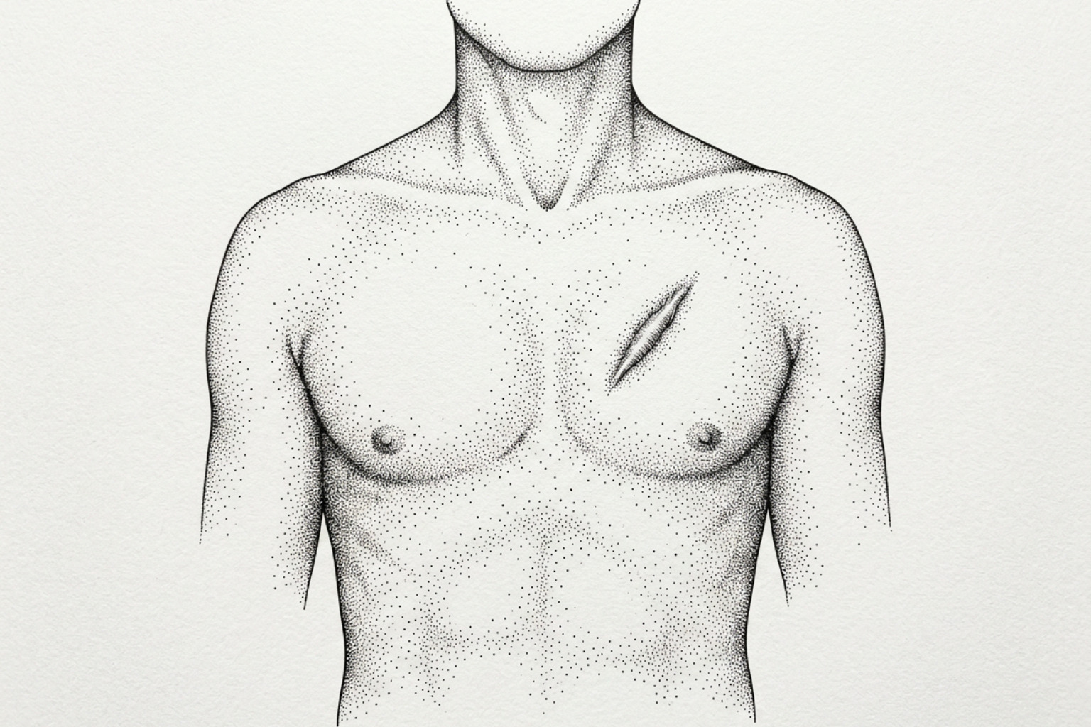
Where a keloid forms matters as much as why. Chest and shoulder keloids sit in high-tension skin and recur far more often than earlobe keloids, and that changes which treatment makes sense.
Where keloids form, and why it matters
Understanding keloid scar body location risk is one of the most practical steps a person can take when managing or preventing these scars — because where a keloid forms matters as much as why it forms.
Quick answer: highest-risk body locations for keloid scars
| Body Location | Risk Level | Notes |
|---|---|---|
| Anterior chest / sternum | Very high | Highest recurrence after treatment (up to 43%) |
| Scapular / upper back | Very high | High skin tension zone |
| Earlobes | High | Common after piercing; lowest recurrence after treatment (~6%) |
| Shoulders | High | Frequent spontaneous keloid site |
| Upper arms / deltoid | Moderate-high | Common post-injection or trauma site |
| Cheeks / jaw | Moderate | Common after acne |
| Abdomen | Moderate | Common after surgery |
| Palms, soles, genitalia | Very low | Rarely affected |
A keloid is not simply a raised scar. It is the result of a dysfunction in the normal wound-healing process, in which fibroblasts — the cells responsible for producing collagen — become overactive. Instead of producing the controlled amount of collagen (a structural protein that makes up the extracellular matrix of skin) needed to close a wound, they continue producing far more than necessary. Clinical data shows collagen synthesis in keloid tissue can run 20 times higher than in healthy skin.
The result is a scar that grows beyond the original wound boundary, invades surrounding healthy tissue, and — unlike a hypertrophic scar — does not regress on its own.
What makes keloids particularly complex is that this process does not happen equally across the body. Certain anatomical sites are far more prone to keloid formation than others, and the same injury — say, a small cut — can heal cleanly on one part of the body while triggering a large keloid on another. Research consistently points to the anterior chest (sternum), scapular region, earlobes, and shoulders as the highest-risk zones, while areas like the palms, soles, and mucous membranes are rarely affected.
This guide maps those location-based risks in detail — covering the biology behind them, how individual factors like skin type, age, and genetics interact with specific body sites, and what the evidence says about prevention and treatment outcomes by location.
Why skin tension drives keloids
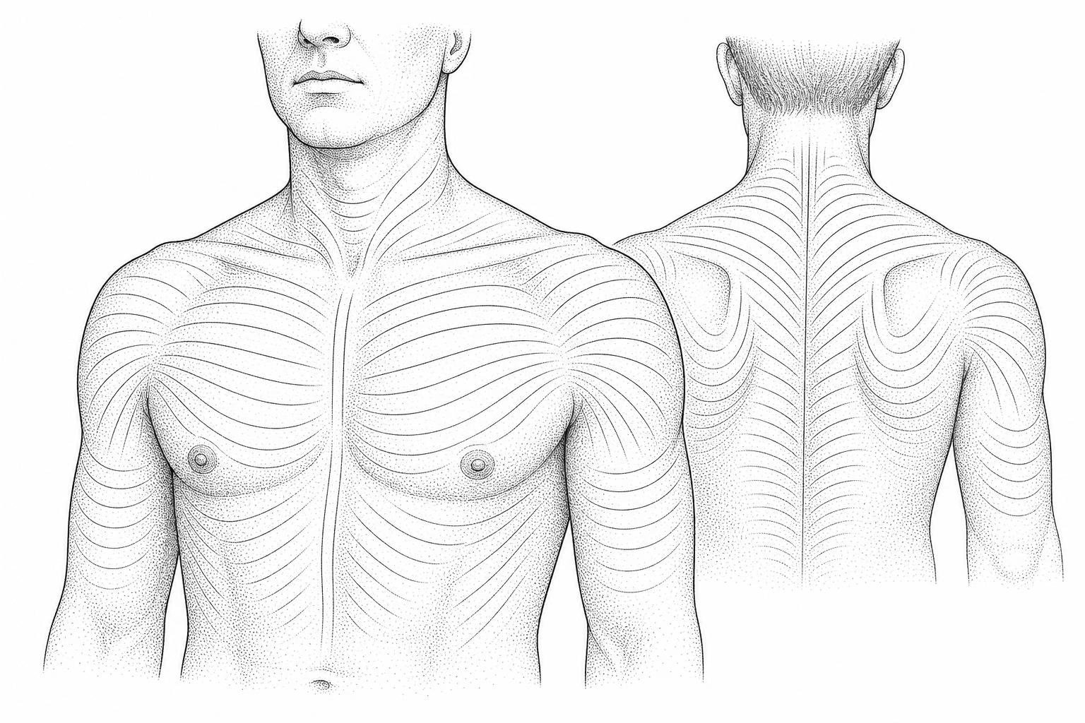
The primary reason keloids show a strong preference for specific anatomical sites comes down to mechanical tension. The human skin is constantly subjected to physical forces from muscle movement, joint flexion, and respiration. Areas like the anterior chest, shoulders, and scapular regions experience high levels of static and dynamic skin stretching.
When an injury occurs in a high-tension zone, the mechanical forces pull at the wound edges. This mechanical stretch is sensed by specialized mechanoreceptors on dermal fibroblasts. In response, these fibroblasts initiate an inflammatory cascade, upregulating transforming growth factor-beta (TGF-β) and other pro-fibrotic cytokines. This chronic inflammatory state prevents the scar from transitioning into the normal remodeling phase of wound healing. Instead, the fibroblasts continue to deposit thick, disorganized collagen bundles (primarily Type I and Type III collagen) into the extracellular matrix.
According to a comprehensive UpToDate clinical review on keloids, these mechanical stretching forces generate a "leading edge" for scar spreading. This explains why keloids in high-tension areas do not remain circular; they expand along the lines of highest skin tension, invading adjacent healthy skin and developing highly characteristic shapes.
Conversely, areas where skin tension is exceptionally low or where the skin is tightly bound to underlying structures without frequent stretching—such as the upper eyelids, the anterior lower leg, and the scalp—rarely develop keloids, even in individuals who are highly predisposed to pathological scarring.
Who gets keloids, and where
While physical tension is a major driver, systemic and genetic factors heavily dictate how a person's body responds to skin trauma. The overall incidence of keloids varies widely among different ethnic populations, ranging from 4.5% to 16% in individuals of African, Asian, and Hispanic descent, compared to less than 0.1% in northern European populations.
This variation is closely tied to melanin density and melanocyte activity. Clinical observations show that keloids are highly prevalent in darkly pigmented skin but virtually non-existent in individuals with albinism. One hypothesis suggests that melanocytes, which are more active in darker skin tones, interact directly with dermal fibroblasts to promote collagen overproduction. Interestingly, keloids are far more likely to occur on body areas with a high density of melanocytes, such as the face and chest, compared to areas with fewer melanocytes, such as the palms and soles.
To understand how these biological factors manifest over time, it is helpful to examine the Keloid Scar Formation Stages, which outline how early inflammation transitions into permanent, self-sustaining fibroproliferative tissue. For a deeper understanding of these underlying genetic and environmental triggers, the StatPearls review on keloid etiology provides an extensive breakdown of how these cellular changes occur.
How age and sex change keloid sites
Age and gender play a significant role in where and when keloids develop. The peak incidence for keloid formation occurs during the second and third decades of life (ages 10 to 30). This period correlates with high rates of epidermal turnover, elevated growth hormone levels, and peak sebum production. Keloids are exceptionally rare in the first decade of life and tend to decrease significantly in older age, suggesting that sexual hormones (estrogen and androgen) play a supportive role in keloidogenesis.
Hormonal fluctuations during puberty and pregnancy are known to accelerate keloid growth and increase the risk of new lesions forming, particularly on the chest, abdomen, and earlobes.
Gender also influences the distribution of keloids, often due to cultural or environmental exposures:
- Females: Historically show a higher overall rate of earlobe keloids, primarily due to the higher prevalence of ear piercing. In pediatric populations, studies show a female-to-male ratio of up to 4.2:1 in childhood-onset cases, frequently linked to early earlobe piercings and vaccinations. For instance, a study on BCG vaccine-induced keloids found that 93% of patients developing keloids at the injection site (typically the deltoid) were female.
- Males: Frequently present with keloids on the cheeks, jawline, and posterior neck (acne keloidalis nuchae). These are often triggered by shaving irritation, folliculitis, or severe cystic acne. In older demographics (above age 40), males show slightly higher rates of trunk and extremity keloids than females, often associated with occupational trauma or surgical procedures.
Genetics and other risk factors
A positive family history is one of the strongest predictors of keloid severity and location. Approximately 30% to 50% of individuals who develop keloids have a first-degree relative with the same condition. Clinical studies indicate that up to 76% of patients with a strong family history develop keloids in the exact same anatomical sites as their relatives, suggesting a highly localized genetic susceptibility.
Beyond family history, several systemic factors correlate with increased risk:
- Blood Group A: Epidemiological data suggests that individuals with blood type A have a statistically higher probability of developing keloids, particularly spontaneous keloids on the sternum and shoulders, compared to other blood groups.
- Genetic Syndromes: Certain genetic conditions, such as Rubinstein-Taybi syndrome, carry an exceptionally high risk, with up to 24% of these patients developing severe, multi-site keloids.
- Hypertension: Elevated blood pressure is positively correlated with both keloid size and the total number of lesions. Chronic hypertension causes microvascular damage and endothelial dysfunction, which sustains a low-grade inflammatory state in the dermis, feeding the overactive fibroblasts in high-tension areas like the chest and scapula.
How the type of injury changes risk
The mechanism of the initial skin injury heavily influences where a keloid is likely to form. Different types of trauma affect different layers of the dermis and carry varying levels of tension:
- Burns: Thermal and chemical burns typically result in deep, widespread dermal damage. Approximately 45% of burn-induced keloids are located on the extremities (both upper and lower limbs), where joint movement exerts continuous mechanical stretching on the healing tissue.
- Piercings: Ear piercings are the single most common cause of keloids on the earlobe and auricle. Because piercing creates a continuous tract of damaged dermis that is slow to epithelialize, it provides an ideal environment for prolonged inflammation and subsequent keloidal growth.
- Acne and Folliculitis: Inflammatory skin conditions are the primary driver of keloids on the face, jawline, chest, and upper back. Acne-related keloids are particularly common on the sternum and shoulders, where skin tension is high and sebaceous gland activity is prominent.
- Surgical Wounds: Surgical incisions, particularly those that run perpendicular to Langer's lines (natural skin tension lines), are highly prone to keloid formation. Approximately 50% of surgical keloids are located on the sternum (following cardiothoracic surgery) and the abdominal wall (following laparotomies or Cesarean sections). For those managing post-operative scarring in the pelvic or abdominal region, specialized protocols such as Abdomen Keloid Scar Reduction offer targeted insights into managing these high-tension surgical scars.
- Spontaneous Keloids: In highly susceptible individuals, keloids can form without any remembered history of trauma. Around 80% of these spontaneous keloids are located on the sternum and shoulders, likely triggered by microscopic, unnoticed micro-trauma, such as friction from clothing or minor folliculitis.
To understand the full range of early symptoms and how various injuries trigger this response, you can consult the Mayo Clinic guide on keloid symptoms and causes.
One keloid vs. keloids in many places
Most individuals with a predisposition to keloids will only develop a single lesion following a specific trauma, such as an ear piercing. However, a subset of the population exhibits "multiple-site" keloid behavior, developing lesions across several distinct anatomical zones.
Studies show that between 19.3% and 42.2% of keloid patients present with multiple-site keloids. This multi-site behavior represents a distinct clinical phenotype characterized by:
- Systemic Susceptibility: These individuals have a much lower threshold for keloid trigger factors. Minor injuries like insect bites, scratches, or vaccinations can trigger large keloids across the body.
- Etiological Patterns: In cohorts with multiple-site keloids, burn injuries are the most common cause (accounting for roughly 38.2% of cases, with the upper limbs being the most common site), followed closely by multiple ear piercings.
- Demographic Risk Factors: Multiple-site keloid behavior is strongly associated with female sex, a younger age at initial presentation, and a positive family history of pathological scarring.
How location affects treatment and recurrence
The anatomical location of a keloid is one of the most critical factors determining how well it will respond to treatment and how likely it is to recur. Treating keloids is notoriously difficult; surgical excision alone carries a recurrence rate of 45% to 100%. However, combining surgery with adjuvant therapies—such as radiation, corticosteroid injections, or silicone sheeting—significantly improves outcomes.
According to a comprehensive Systematic review on keloid treatment efficacy, keloids located on the chest, shoulders, and extremities demonstrate much lower treatment efficacy and higher recurrence rates compared to those on the face or earlobes. High-tension zones keep the wound in a prolonged inflammatory state, which continuously stimulates fibroblasts even after the bulk of the scar has been removed or treated.
To navigate the wide variety of treatment pathways, patients and clinicians often look to options like Laser for Keloid Scars, Non-Invasive Keloid Removal, and various methods of Keloid Treatment Without Surgery to minimize the risk of triggering a larger recurrence through surgical trauma.
| Body Location | Primary Causes | Recurrence Risk | Recommended First-Line Treatments |
|---|---|---|---|
| Earlobe | Piercings, trauma | Low (~5% - 12%) | Wedge/core excision + pressure earrings + low-dose radiation (8 Gy) |
| Anterior Chest | Surgery, acne, spontaneous | Very High (34% - 43%) | Tension-free excision + immediate postoperative radiation (18 Gy) + silicone sheeting |
| Shoulders / Scapula | Acne, vaccines, trauma | Very High (30% - 40%) | Corticosteroid injections + laser therapy + long-term silicone tape |
| Face (Cheeks / Jaw) | Acne, shaving, trauma | Moderate (15% - 25%) | Intralesional steroids + 5-FU + pulsed dye laser (PDL) |
| Abdomen | Surgery (C-sections, laparotomy) | Moderate (20% - 30%) | Surgical revision with tension-reduction sutures + silicone gel sheeting |
| Extremities | Burns, trauma, surgery | Moderate-High (25% - 35%) | Compression garments + corticosteroid injections + silicone sheets |
Treatment options by body area
Because the biological environment varies so much by body part, clinical protocols must be customized by location to be effective. The Merck Manual professional reference on keloids outlines standard clinical approaches, which are typically adapted as follows:
- Earlobes: These are the easiest keloids to treat successfully. Because the earlobe is a low-tension zone, the recurrence rate after customized treatment is only 5% to 7%. The standard protocol involves a wedge or core excision to preserve the shape of the ear, followed immediately by a single dose of postoperative radiotherapy (8 Gy) or the use of mechanical pressure earrings worn for 20 hours a day for six months.
- Anterior Chest Wall: This is the most difficult region to treat, with recurrence rates historically exceeding 40%. Because the chest is constantly moving due to respiration, surgical wounds are under constant tension. The current gold-standard protocol requires a highly specialized surgical technique using deep fascial tension-reduction sutures to take the strain off the skin surface. This must be followed by immediate postoperative electron-beam radiation (typically 18 Gy split over three days, starting the day after surgery) and continuous stabilization with silicone sheets for at least six months.
- Face and Neck: Protecting aesthetics is the primary concern here. Surgical excision is often avoided as a first step due to the risk of creating a larger scar. Instead, clinicians prefer non-surgical combination therapies, such as monthly intralesional corticosteroid injections (triamcinolone acetonide) combined with 5-fluorouracil (5-FU) and pulsed dye laser (PDL) sessions to reduce vascularity and flatten the scar.
Preventing keloids in high-risk areas
For individuals with a known predisposition to keloids, primary prevention is far more effective than trying to treat a fully formed scar. When skin trauma occurs in high-risk zones, implementing early intervention protocols can prevent the overactivation of fibroblasts.
According to the StatPearls guide on hypertrophic scarring and keloids, several evidence-based preventive measures should be taken:
- Avoid Elective Trauma: If you have a personal or strong family history of keloids, avoid elective procedures such as body piercings, tattoos, and non-essential cosmetic surgeries, especially on high-risk areas like the chest, shoulders, and ears.
- Ensure Rapid Wound Healing: Keep any wound clean, moist, and covered. Using petrolatum jelly (Vaseline) to keep the wound environment hydrated accelerates re-epithelialization. The faster a wound heals, the less time it spends in the inflammatory phase that triggers keloid growth.
- Apply Mechanical Stabilization: Immediately after a wound has closed (usually 2 to 3 weeks post-injury), apply silicone gel sheeting. Silicone sheets help hydrate the stratum corneum and transmit subtle mechanical signals that downregulate fibroblast activity. In high-tension areas, use paper tape or silicone tape to physically hold the skin together, reducing the lateral tension on the immature scar.
- Utilize Compression Therapy: For extremity and earlobe injuries, applying continuous pressure (between 15 to 45 mm Hg) for at least 20 hours a day for 3 to 6 months helps restrict local blood flow. This limits the supply of oxygen and nutrients to the overactive fibroblasts, slowing down collagen synthesis.
Frequently asked questions
What are the rarest body locations for keloid scars?
The rarest body locations for keloid scars include the palms of the hands, the soles of the feet, the genitalia, the buttocks, and mucous membranes (such as the inside of the mouth). These areas have very low rates of mechanical stretching, lack sebaceous glands, and have a unique dermal structure with lower melanocyte activity, making them highly resistant to pathological scarring.
Why do chest keloids often form in a butterfly or crab-claw shape?
Chest keloids often form in a butterfly or "crab-claw" shape because they grow along the lines of highest physical tension. On the anterior chest, the skin is pulled horizontally by the major pectoral muscles and vertically by the weight of the torso and respiratory movements. As the keloid expands, it follows these prevailing tension lines, spreading outward into healthy tissue while leaving the lower-tension center relatively spared.
Can keloids form spontaneously without a known skin injury?
Yes, spontaneous keloids can form on uninjured skin, most commonly on the sternum and shoulders. However, these are rarely truly "spontaneous." Instead, they are typically triggered by microscopic, unnoticed dermal inflammation, such as a minor insect bite, a blocked hair follicle (folliculitis), friction from clothing, or a tiny scratch that occurred months prior.
Key takeaways
Managing pathological scarring requires a deep understanding of how physical location shape-shifts the behavior of a scar. By identifying high-risk zones like the chest and shoulders early, individuals and clinicians can make highly informed decisions regarding preventive care, surgical planning, and targeted therapies.
For those looking to evaluate their personal risk or seek professional guidance on an existing scar, utilizing specialized scar assessment services is an excellent next step toward finding a personalized, clinically proven path to healing.
Works Cited
- Bekkers, V., Barsoum, P., Yin, Q., Niessen, F., van Zuijlen, P., Lapid, O., van Doorn, M. & Wolkerstorfer, A. "Effect of Keloid Properties on Treatment Efficacy: A Systematic Review." Dermatologic Surgery, vol. 50, no. 10, 2024, pp. 913–921.
- Ogawa, R. "Keloid and Hypertrophic Scars Are the Result of Chronic Inflammation in the Reticular Dermis." International Journal of Molecular Sciences, vol. 18, no. 3, 2017, art. 606.
- Carswell, L. & Borger, J. "Hypertrophic Scarring and Keloids." StatPearls, NCBI Bookshelf NBK537058, 2023.
- McGinty, S. & Siddiqui, W. J. "Keloid." StatPearls, NCBI Bookshelf NBK507899 (archived edition, retained for historical reference).
- "Keloids and Hypertrophic Scars." UpToDate (subscription required).
- "Keloid Scar — Symptoms and Causes." Mayo Clinic.
- "Keloids." Merck Manual Professional Edition.
This content is for informational purposes only and does not constitute medical advice. Consult a qualified healthcare professional for diagnosis and treatment.