The Great Debate of Scar Gels vs Creams in Modern Protocols
Dermatology protocols favor silicone gels and sheets as first-line scar treatment. This guide compares clinical evidence for gels, sheets, lasers, and injections across every healing stage.
What the Research Shows
Dermatology scar management protocols refer to the structured, evidence-based approaches clinicians use to prevent, monitor, and treat abnormal scar formation following surgery, trauma, or inflammatory skin conditions.
Quick reference — core protocol steps by scar stage:
| Stage | Timing | Key Interventions |
|---|---|---|
| Prevention | During/immediately after surgery | Incision design, tension reduction, moist wound healing |
| Early intervention | 2–4 weeks post-wound closure | Silicone gel or sheets, taping, sun protection |
| Active management | 4 weeks – 6 months | Corticosteroid injections, pressure therapy, laser |
| Refractory/pathological | 6–12+ months | 5-FU, cryosurgery, surgical revision, radiotherapy |
| Long-term monitoring | Up to 24 months | Re-evaluation, combination therapy, psychosocial support |
Scars are not a single problem with a single solution. Skin heals through three overlapping phases — inflammation, proliferation, and remodeling — and what happens in each phase determines the final appearance of a scar. When that process goes wrong, the result can be a raised keloid, a sunken atrophic depression, or a wide hypertrophic band across the skin.
The scale of this issue is significant. Research estimates that around 100 million people in the developed world acquire scars each year following surgery alone, and roughly 15% of those develop scars that are excessive or aesthetically distressing. A separate survey found that 91% of surgical patients would value any measurable improvement in their scarring.
Despite the clinical demand, there is still no single gold-standard treatment algorithm. The pathophysiology of scarring is complex, individual responses vary considerably, and many popular products have limited rigorous clinical evidence behind them. This guide examines what the current evidence actually supports — from silicone gels and sheets to laser therapy and adjunctive injections — and where the science still has gaps.
How Scars Are Classified
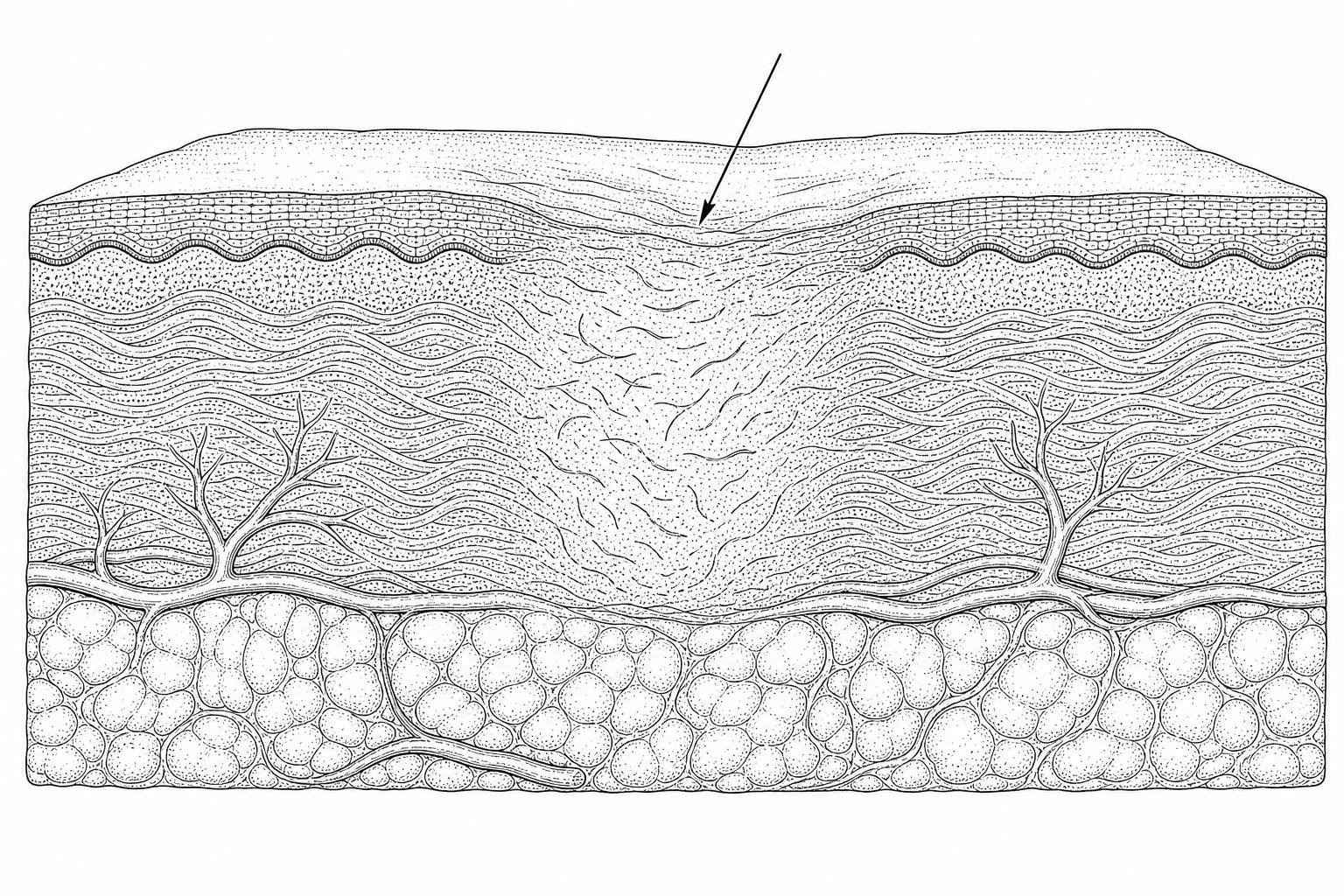
Understanding the biological mechanisms of wound healing is essential for effective scar assessment. When the deep dermis is injured, the body initiates a repair cascade involving hemostasis, inflammation, proliferation, and maturation. Fibroblasts, the primary cells responsible for tissue repair, migrate to the site to synthesize a new extracellular matrix (ECM).
In normal healing, collagen production and degradation eventually reach an equilibrium. However, dysregulation in this process leads to pathological scarring. Hypertrophic scars and keloids both result from excessive fibroblast activity and collagen deposition, but they differ significantly in their clinical behavior.
- Hypertrophic Scars: These are raised, red, and firm lesions that remain within the boundaries of the original wound. They often develop within weeks of injury and may partially regress over one to two years. Histologically, they are characterized by well-organized bundles of collagen type III.
- Keloids: These represent a more aggressive fibrotic disorder. Keloids grow beyond the margins of the original injury, invading surrounding healthy skin. They rarely regress spontaneously and contain disorganized, thick bundles of collagen types I and III. Understanding the biological stages of keloid formation can help identify when to intervene.
- Atrophic Scars: These are characterized by a loss of tissue, resulting in depressions or "pitted" skin. They commonly follow inflammatory conditions like acne or varicella (see our chickenpox scar treatment guide for adults). Management often requires specialized approaches such as atrophic scar microneedling therapy.
How Scar Protocols Have Evolved
Recent shifts in clinical thinking emphasize moving beyond visible phenotypes (what the scar looks like) to understanding endotypes (the underlying pathophysiological mechanisms). A proposed framework for this precision approach uses the acronym S.C.A.R.: Stretched, Contracted, Atrophic, and Raised.
This evolution in dermatology scar management protocols also highlights the role of mechanotransduction—how mechanical forces on a wound are converted into cellular signals. High skin tension is a primary driver of fibroblast proliferation. The National expert consensus on early management of scars suggests that intervening early, while the scar is still immature, can significantly improve long-term outcomes by modulating these mechanical and biological signals before they become permanent.
Patient Factors Influencing Scar Outcomes
Clinical outcomes are heavily influenced by unmodifiable patient factors. Genetics and ethnicity play a major role; for instance, populations of African, Asian, and Hispanic descent experience a higher incidence of keloids, with rates as high as 4.5% to 16% in some groups.
Age also dictates healing patterns. Children are more prone to hypertrophic scarring due to higher skin elasticity and rapid physical growth, which increases wound tension. Conversely, the elderly often form finer scars due to decreased skin tension and cellular activity. Anatomical location is equally critical; areas of high movement or tension, such as the chest, shoulders, and joints, are high-risk zones for pathological scarring. For younger patients, specialized guidance like a pediatric scar reduction cream guide is often necessary to account for these developmental differences.
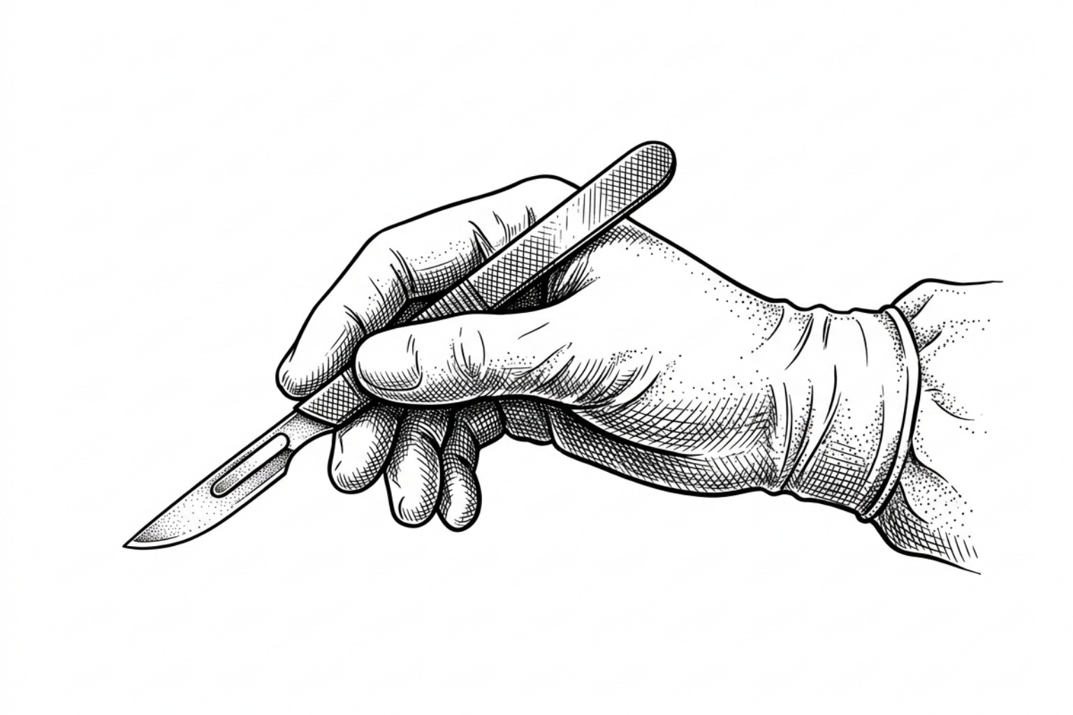
Prevention Starts in the Operating Room
The foundation of scar prevention begins in the operating room. Expert consensus indicates that the single most important modifiable factor in scar prevention is wound tension. Surgeons utilize Relaxed Skin Tension Lines (RSTLs)—the natural folds of the skin—to guide incision design.
Clinical guidelines emphasize the "5 A's" of surgical technique: asepsis, absence of tension, accurate approximation, avoidance of raw surfaces, and atraumatic tissue handling. Proper post surgery scar care continues this focus on stability. At one week post-surgery, an incision has only 3% of the tensile strength of normal skin, reaching 20% by week three and 80% after three months. Understanding when to apply scar cream after stitches is vital; most protocols recommend waiting until the wound has fully epithelialized and sutures are removed.

Immediate Post-Surgery Care
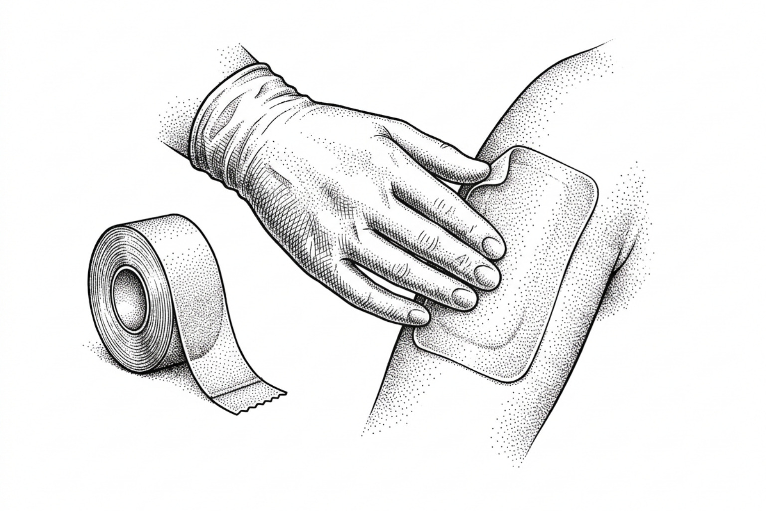
The goal immediately after wound closure is to ensure rapid epithelialization (ideally within 10–14 days) and maintain a moist environment. Delayed healing is a known risk factor for hypertrophic response. Polyurethane dressings and hypoallergenic microporous tape are frequently used to offload tension from the wound edges. For specific surgical recoveries, such as a non-invasive C-section scars guide, these taping techniques are often maintained for up to 12 weeks to provide continuous support during the early remodeling phase.
Long-Term Monitoring and Remodeling Phases
The remodeling phase of a scar can last up to 24 months. During this window, the scar's vascularity decreases, and collagen fibers reorganize. A standard 12-month monitoring timeline is recommended, with clinical re-evaluations every 4 to 8 weeks in the early stages. If a scar remains erythematous (red) or "upset" beyond the initial months, clinicians may introduce hard scar softening massage techniques or more aggressive interventions to prevent permanent hypertrophy.
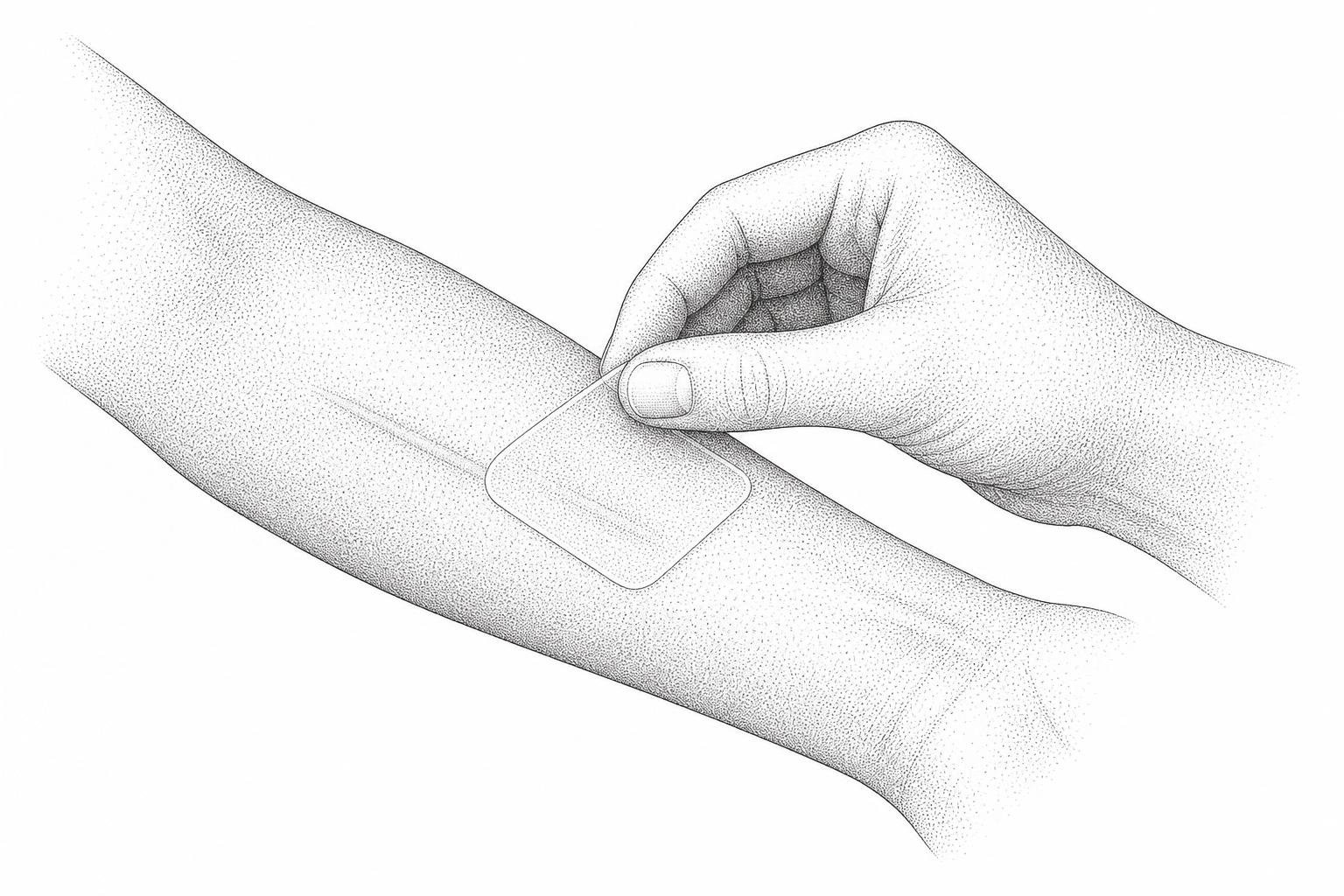
Silicone Gel vs. Silicone Sheets
Silicone-based products remain the gold standard for non-invasive scar management. Their primary mechanism of action is not through "absorption" of silicone, but through occlusion and hydration. By mimicking the skin's barrier, silicone reduces transepidermal water loss (TEWL), which signals fibroblasts to slow down collagen production.
| Feature | Silicone Gel Sheets | Liquid Silicone Gels |
|---|---|---|
| Best For | Large scars, flat areas, overnight use | Visible areas (face), joints, children |
| Mechanism | Occlusion + slight pressure | Occlusion via self-drying film |
| Wear Time | 12–24 hours/day | Twice daily application |
| Pros | High efficacy, reusable | Discrete, easy to apply |
| Cons | Visible, can cause skin irritation | Requires drying time (4–5 mins) |
The Updated International Clinical Recommendations on Scar Management confirm that both formats are effective, though sheets may offer slightly superior results due to the added benefit of minor pressure. However, patient compliance is the ultimate deciding factor. For those struggling with raised tissue, reviewing hypertrophic scar silicone gel evidence can help in selecting the most appropriate format for their lifestyle.
Efficacy of Topical Formulations and Over-the-Counter Agents
While silicone is the gold standard, many patients seek over-the-counter (OTC) alternatives. Choosing among them can be daunting — our best silicone scar cream comparison ranks the top options. The evidence for these is often mixed:
- Onion Extract: Some studies suggest a modest improvement in scar redness and texture when applied twice daily for 3 to 6 months.
- Vitamin E: Despite its popularity, Vitamin E scar healing research shows inconsistent results, with some studies indicating no benefit and a significant risk of contact dermatitis (skin rash).
- Aloe Vera: This is often used for its anti-inflammatory properties. While an Aloe vera scar reduction guide may suggest benefits for soothing fresh scars, it is generally considered an adjunctive treatment rather than a primary scar-remodeling agent.
- Green Tea (EGCG): Emerging research suggests that pre-treating skin with green tea extract before surgery may help reduce subsequent scar formation by inhibiting certain pro-fibrotic cytokines. For product comparisons, see the top-rated scar reduction creams.
Future Directions in Scar Science
The future of scar management lies in precision medicine. Researchers are investigating biomarkers that can predict which patients are most likely to develop keloids. New therapies targeting specific molecular pathways, such as mechanotransduction inhibitors (e.g., FAK inhibitors), are currently under exploration. The goal is to move beyond managing existing scars toward achieving "scarless" healing through the modulation of the inflammatory environment.
Treatments for Stubborn or Severe Scars
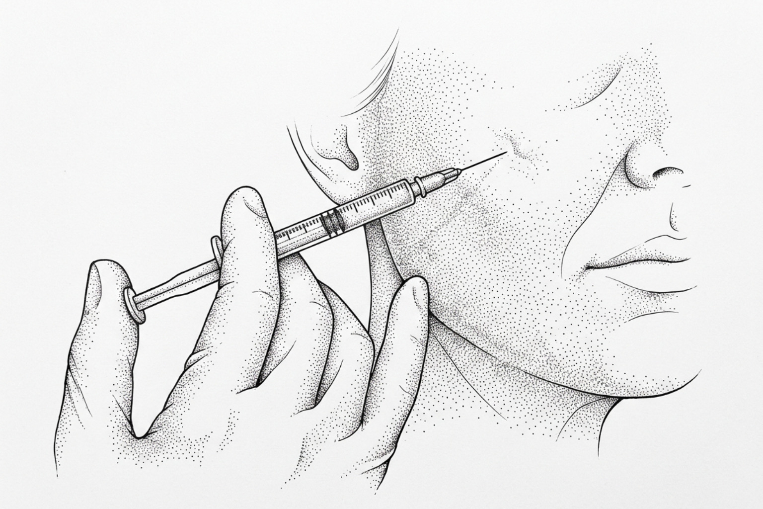
When non-invasive methods like silicone fail, clinicians turn to intralesional therapies. These involve injecting medications directly into the fibrous tissue of the scar to break down excess collagen.
- Corticosteroids: Triamcinolone acetonide (TAC) is the most common injection. It reduces inflammation and inhibits fibroblast growth. Research shows TAC can reduce the size of raised scars by 50% or more.
- 5-Fluorouracil (5-FU): This chemotherapy agent is increasingly used for keloids that don't respond to steroids. It inhibits fibroblast proliferation. Evidence suggests that a combination of 5-FU and TAC is often more effective than either alone, with lower recurrence rates.
- Bleomycin: This antibiotic with anti-tumor properties is used for refractory scars, showing high success rates in flattening stubborn keloids.
For those seeking to avoid surgery, exploring non-invasive keloid removal options such as cryosurgery is often the next step. Cryosurgery involves freezing the scar tissue from the inside out, which can reduce scar volume by over 50% after a single session.
The Role of Laser Therapy and Surgical Revision
Laser therapy has become a cornerstone of modern dermatology scar management protocols. For a deeper comparison of laser technologies, see our complete laser treatment guide. Different lasers target different aspects of the scar:
- Pulsed Dye Laser (PDL): Targets the blood vessels in the scar to reduce redness and itching. Learn more about laser treatments for red scars.
- Fractional CO2 Laser: Creates microscopic "holes" in the scar to stimulate the production of new, more organized collagen. This is particularly effective for CO2 laser for surgical scars before and after treatments.
- Ablative Lasers: Used to "resurface" the skin, smoothing out the texture of hypertrophic or laser for keloid scars.
If a scar is functionally limiting or aesthetically unacceptable after 12 months, a surgical scar revision complete guide may be consulted. Techniques like Z-plasty or W-plasty are used to reposition the scar or break up linear tension, making the final result less noticeable.
Preventing Scars from Coming Back
Because keloids have a high recurrence rate after surgery (45% to 100%), surgeons almost always use adjunctive therapies. Radiation therapy, specifically electron beam irradiation, is often administered within 24–48 hours of surgical excision to prevent new keloid tissue from forming.
Pressure therapy is another vital tool, especially for burn survivors. For a broader overview of treatment options, see the best burn scar treatment guide. By applying constant pressure (15–40 mmHg) for 23 hours a day, clinicians can induce fibroblast apoptosis (cell death) and reduce collagen synthesis. This is often paired with scar tissue massage therapy to maintain tissue pliability during the long maturation process.
Common Questions About Scar Management
How Long to Use Silicone Therapy
Clinical guidelines recommend using silicone gel or sheets for a minimum of 12 hours a day (ideally 24 hours) for at least 3 to 6 months. Maximum improvement is typically seen after 2 to 3 months of consistent use.
Optimal Timing for Laser Intervention
While traditionally doctors waited for a scar to "mature," modern protocols often support early intervention. Vascular lasers (PDL) can be used as soon as 2 to 4 weeks after suture removal to manage redness, while fractional lasers may be introduced within the first few months to optimize remodeling.
Efficacy of Natural Oils in Scar Management
There is limited high-level clinical evidence that natural oils like coconut or olive oil significantly change the structure of a scar. However, the act of massaging the oil into the skin can help keep the tissue hydrated and pliable, which may improve comfort and appearance.
Building Your Scar Treatment Plan
Managing scars effectively requires an individualized, multidisciplinary approach that begins the moment an injury occurs. By combining surgical precision with evidence-based non-invasive treatments like silicone and advanced interventions like lasers, clinicians can significantly improve both the functional and aesthetic outcomes for patients. For those beginning their journey, utilizing a complete scar assessment tool is the first step toward a personalized treatment plan.
Works Cited
- Gold, M. H., et al. (2014). "Updated International Clinical Recommendations on Scar Management: Part 2—Algorithms for Scar Prevention and Treatment." Dermatologic Surgery, 40(8), 825–831.
- Mustoe, T. A., et al. (2002). "International Clinical Recommendations on Scar Management." Plastic and Reconstructive Surgery, 110(2), 560–571.
- Basson, R. & Bayat, A. (2022). "Skin Scarring: Latest Update on Objective Assessment and Optimal Management." Frontiers in Medicine, 9, 942756.
- Sequeira Campos, M. B., Xiao, A. & Ettefagh, L. (2025). "Laser Revision of Scars." StatPearls. Treasure Island, FL: StatPearls Publishing.
- Zheng, Y., et al. (2021). "National Expert Consensus on Early Management of Scars (2020 Version)." Chinese Journal of Burns, 37(2), 113–125.
This content is for informational purposes only and does not constitute medical advice. Consult a qualified healthcare professional for diagnosis and treatment.