Steroid injection versus silicone and pressure: how clinicians choose.
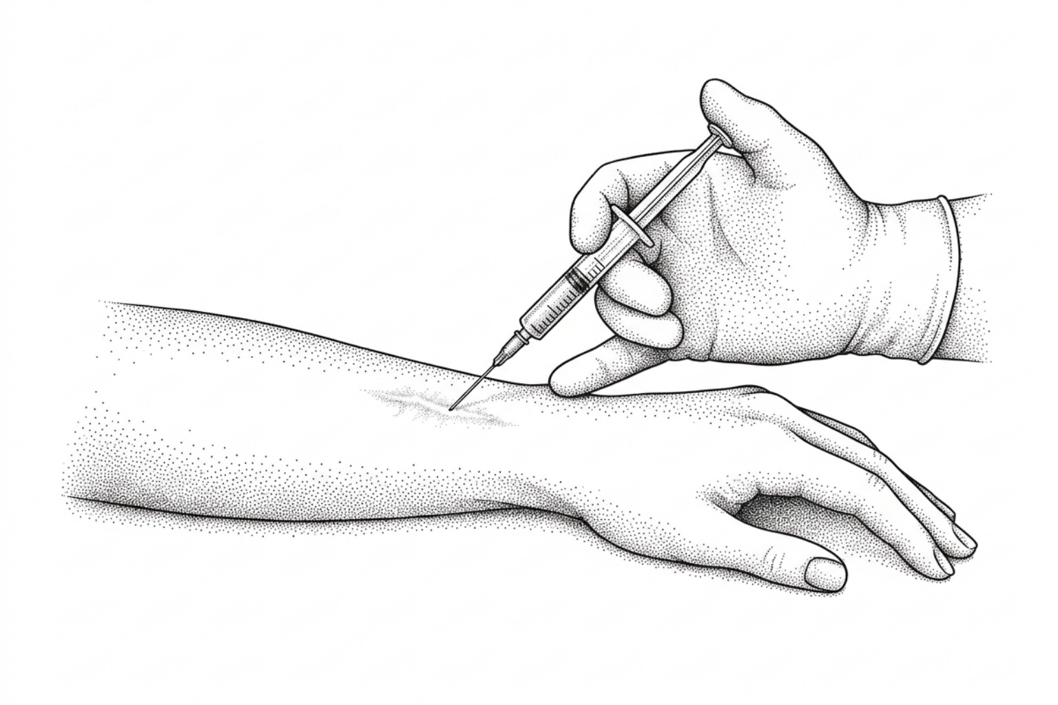
Steroid injections, silicone sheeting and pressure therapy all flatten raised scars, but they suit different scars and different lives. Here is how clinicians choose between them, and what the trial evidence actually shows.
How keloids and hypertrophic scars form
When evaluating steroid injection versus silicone and pressure, clinicians primarily choose based on scar maturity, tissue thickness, and biological boundaries. Non-invasive silicone gel sheeting serves as the preferred first-line intervention for immature hypertrophic scars and surgical prophylaxis by restoring stratum corneum hydration. In contrast, intralesional corticosteroid injections are indicated for dense, rigid keloids and established hypertrophic scars requiring rapid collagen degradation and volumetric reduction. Pressure therapy is predominantly reserved for extensive burn hypertrophic scarring and specific anatomical sites such as earlobes where sustained mechanical occlusion can be maintained.
Abnormal scarring represents a disruption in the physiological wound healing cascade, which progresses through inflammatory, proliferative, and tissue remodeling phases over 6 to 24 months. During standard healing, fibroblasts synthesize extracellular matrix components that gradually organize into mature bundles. When regulatory signaling fails, sustained activation of transforming growth factor-beta (specifically isoforms TGF-β1 and TGF-β2) leads to uninhibited fibroblast proliferation and excessive collagen deposition.
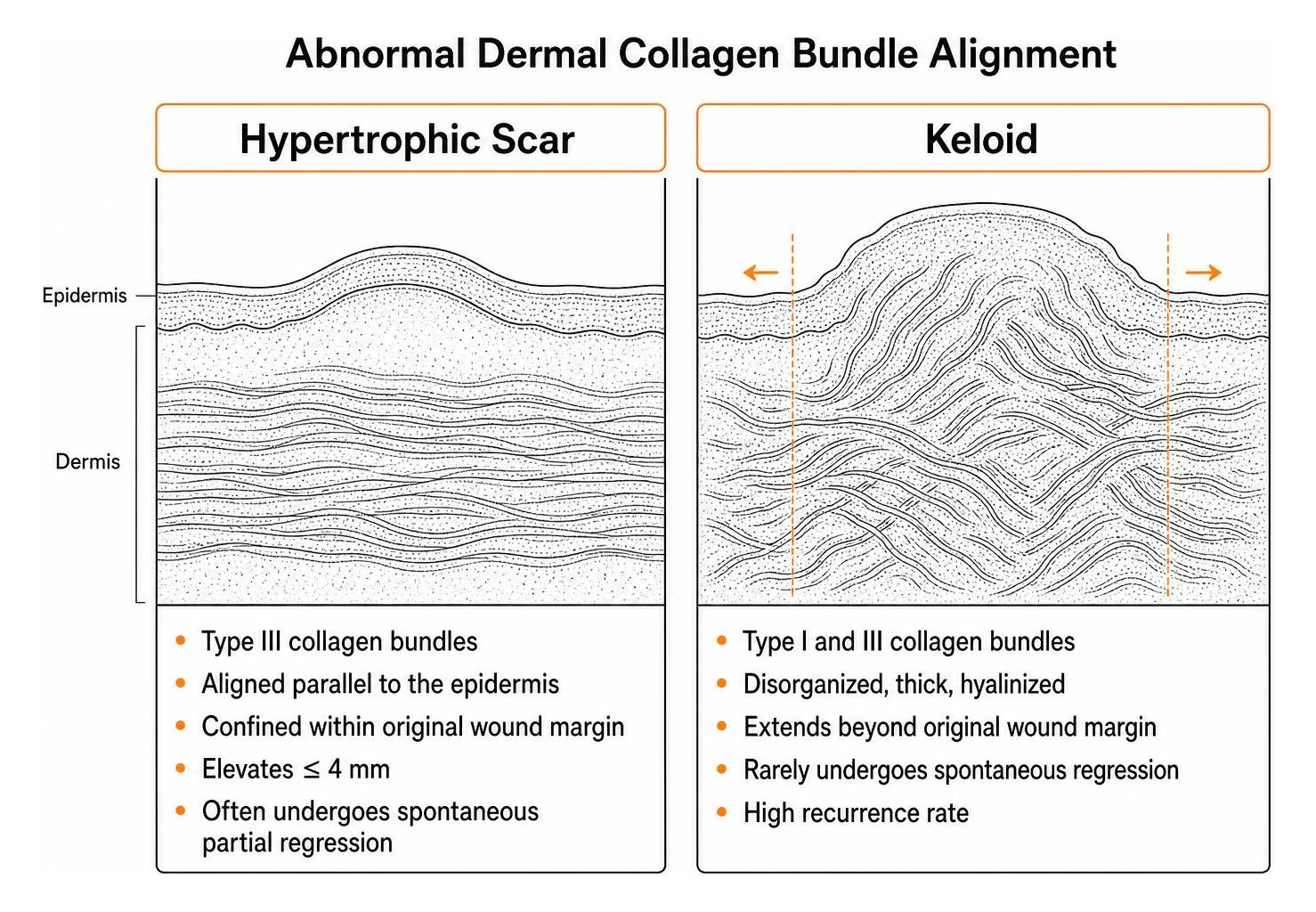
Clinicians distinguish between two primary forms of excessive fibroproliferation:
- Hypertrophic Scars: These lesions arise following thermal injury, surgical incisions, or trauma, with reported incidences ranging from 30% to 91% after severe burns and 39% to 68% after surgical procedures. Histologically, hypertrophic scars contain predominantly type III collagen bundles aligned parallel to the epidermis. They remain strictly confined within the boundaries of the original wound margin, rarely elevate more than 4 mm, and frequently undergo spontaneous partial regression as cellular water content normalizes.
- Keloids: In contrast, keloids represent a progressive dermal proliferation that extends beyond the original margins into adjacent healthy skin. Keloid collagen architecture is characterized by thick, hyalinized, disorganized type I and type III collagen bundles lacking lymphatic channels. Keloids rarely regress spontaneously, demonstrate high recurrence rates, and occur more frequently in darker skin tones (with a 15- to 20-fold increased risk in individuals of African, Asian, or Hispanic descent) and in high-tension anatomical regions such as the presternum, deltoids, and upper back.
Selecting an appropriate treatment for keloid scars begins with verifying this histological and clinical boundary, as true keloids demonstrate much higher biological resistance to non-invasive therapies than immature hypertrophic lesions.
How the three treatments compare
Clinicians evaluating non-operative approaches often contrast intralesional pharmacological therapies with non-invasive mechanical or occlusive modalities. When evaluating steroid injection versus silicone and pressure, published trials reflect starkly different methodologies, study populations, and evidentiary strengths.
| Modality | Level of Evidence (Oxford CEBM) | Primary Mechanism | Reported Response Rate | Common Clinical Indications | Key Practical Limitations |
|---|---|---|---|---|---|
| Intralesional Corticosteroid (TAC) | Level 4 (primarily observational cohorts and small comparative series) | Fibroblast inhibition, collagenase activation, vasoconstriction | 50% – 100% (Recurrence: 9% – 50%) | Rigid, symptomatic keloids; mature hypertrophic scars; adjuvant post-excision care | Procedural pain; dermal atrophy; telangiectasia; pigment alteration (up to 63%) |
| Silicone Gel Sheeting (SGS) | Strong for surgical and burn scars, Moderate for hypertrophic (CEBM Level 2, randomized trials and systematic reviews) | Stratum corneum hydration, reduced TEWL, cytokine normalization | 50% – 100% (Variable magnitude) | Linear surgical scars, immature hypertrophic scars, burn rehabilitation | Requires 12–24 hr/day for 2–3 months; skin maceration; adhesive failure |
| Pressure Therapy | Moderate (CEBM Level 2-3, randomized trials in burn cohorts) | Mechanical hypoxia, fibroblast apoptosis, MMP-28 modulation | 60% – 85% (Burn literature) | Extensive burn hypertrophic scars, earlobe lesions (pressure clips) | Requires 24–30 mm Hg for 6–12 months; garment heat, discomfort, low compliance |
While intralesional triamcinolone acetonide (TAC) has served as a dermatological standard since the 1960s, formal trials evaluating it often sit at Level of Evidence 4 due to lack of blinding, variable dosing, and small cohort sizes. Conversely, topical silicone dressings carry Level of Evidence 2 support, though systematic reviews highlight high risks of detection and performance bias across published studies. For patients seeking keloid treatment without surgery, these evidentiary nuances dictate whether an invasive or non-invasive initial pathway is indicated.
If you have a hypertrophic scar
In hypertrophic scarring, non-invasive topical interventions are generally prioritized before escalating to invasive injections. The therapeutic mechanism of silicone gel sheeting does not rely on pressure, heat transfer, or silicone absorption into dermal layers. Research indicates that medical-grade silicone acts by creating an occlusive barrier that decreases the transepidermal water loss (TEWL) rate to roughly half that of unshielded skin. This continuous hydration mimics a healthy stratum corneum, signalling basal keratinocytes to downregulate pro-inflammatory cytokines that stimulate underlying dermal fibroblasts.
A Cochrane systematic review evaluating 20 randomized controlled trials (873 participants) found that silicone gel sheeting produced a statistically significant reduction in scar thickness compared to untreated controls (mean difference: -2.00 mm; 95% CI: -2.14 to -1.85) alongside improvements in scar pigmentation (risk ratio: 3.49; 95% CI: 1.97 to 6.15). In prevention studies on high-risk individuals, silicone sheeting reduced the incidence of hypertrophic scarring (risk ratio: 0.46; 95% CI: 0.21 to 0.98). However, reviewers emphasized that these trials were highly susceptible to bias, primarily due to non-blinded assessors and unstandardized compliance tracking.
When conservative topical options like hypertrophic scar silicone gel are insufficient for thick, symptomatic hypertrophic scars, intralesional corticosteroids offer rapid matrix breakdown. Corticosteroids arrest collagen synthesis by decreasing alpha-globulin levels (a major inhibitor of natural human collagenase) and suppressing fibroblast mitosis. To minimize extra-tissue extravasation and adverse local changes, clinicians are increasingly exploring guided procedural precision, drawing on technique principles documented in ultrasound versus palpation‐guided corticosteroid injection research to ensure medication remains precisely deposited within dense connective architecture.
If you have a keloid
True keloids exhibit a rigid extracellular matrix with high internal tissue pressure, making them less responsive to topical hydration alone. Head-to-head clinical trials illustrate significant differences in volumetric reduction between these modalities:
- Efficacy in Scar Volume: In a randomized comparative trial by Tan (1999), 94% of participants (16 of 17) treated with intralesional triamcinolone acetonide (40 mg/mL) achieved a greater than 50% reduction in keloid volume, compared to only 12% (2 of 17) in the silicone gel sheeting group (RR: 0.13; 95% CI: 0.03 to 0.46).
- Symptom Onset and Tolerability: Conversely, Sproat et al. (1992) demonstrated that patients using silicone gel sheeting experienced faster symptomatic relief from pain and pruritus (mean difference: -2.90 days; 95% CI: -3.93 to -1.87) and expressed a higher overall treatment preference (RR: 5.50; 95% CI: 1.48 to 20.42) compared to those receiving painful triamcinolone injections.
- Physical Scar Firmness: In a prospective 6-month randomized study on sternotomy scars, researchers utilized mechanical durometers alongside the modified Vancouver Scar Scale (VSS) to objectively track tissue pliability. Steroid monotherapy and combination therapy (steroid plus silicone) achieved statistically significant reductions in durometer-measured tissue hardness (p = 0.044 and p = 0.023, respectively), whereas silicone sheeting alone did not reach statistical significance for tissue softening over the 6-month evaluation.
The physical delivery of intralesional corticosteroids into dense keloidal tissue presents mechanical challenges. Research by Hayward et al. on pressure generated by syringes in connective tissue lesions demonstrated that fluid pressure generation is inversely proportional to syringe barrel cross-sectional area. A 1 mL syringe generates a mean pressure of 363 ± 197 psi (with theoretical peaks up to 600 psi), achieving a 100% success rate in dense tissue hydrodissection, whereas a 10 mL syringe generates only 53 ± 29 psi and fails in 66% of dense lesion attempts. Inadequate injection pressure leads to extralesional drug extravasation into normal subcutaneous fat, precipitating localized tissue atrophy rather than keloid resolution. For refractory lesions, regimens such as corticosteroid injection combined with 5-FU are increasingly utilized to suppress collagen synthesis while allowing lower corticosteroid doses.
Side effects and daily commitment
Every scar management modality carries distinct clinical trade-offs between biological efficacy and patient tolerability:
Steroid injections
The primary limitation of intralesional triamcinolone acetonide (10 to 40 mg/mL) is procedural pain and localized tissue toxicity. Up to 63% of treated lesions develop adverse secondary changes, including cutaneous atrophy, telangiectasias, and permanent hypopigmentation (due to melanocyte suppression). Sproat (1992) reported that 71% of patients experienced severe procedural pain and 64% exhibited localized pigmentary changes or white crystalline deposits following steroid injections, compared to a single instance of mild superficial rash in the silicone group.
Silicone Gel Sheeting
Silicone carries a low risk profile, primarily limited to contact dermatitis, superficial miliaria, and skin maceration. These issues occur primarily when sheets are worn in hot, humid environments or applied without daily washing. In pediatric cohorts, compliance with silicone gel monotherapy remains high (reported at 91.4% in randomized burn trials).
Pressure Therapy
Compression therapy relies on maintaining a continuous mechanical force of 24 to 30 mm Hg for 6 to 12 months to induce localized capillary hypoxia and fibroblast apoptosis. However, clinical adherence is frequently poor due to garment tightness, heat retention, friction blisters, and skin breakdown.
A multicentre randomized controlled trial by Wiseman et al. evaluated 153 pediatric burn patients and challenged the assumption that multi-modal therapy is universally superior. The trial demonstrated that combining silicone gel with pressure garments significantly reduced adherence (silicone adherence fell from 91.4% to 82.8%; pressure adherence dropped from 77.5% to 58.5%) without providing any statistically significant or clinically meaningful improvement in scar thickness or pruritus compared to monotherapy. In addition, pressure garment therapy alone caused significantly higher rates of skin breakdown and mechanical friction injuries than topical silicone.
Combining treatments, and when to start
Clinical guidelines suggest structuring scar management around wound maturity, anatomical location, and recurrence risk:
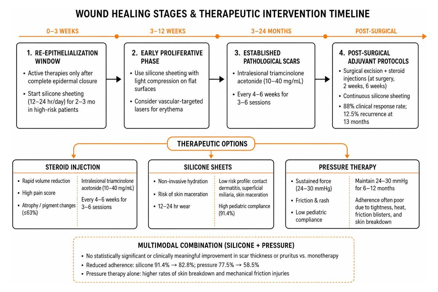
- Re-Epithelialization Window (Weeks 0–3): Active scar therapies should never be applied to open wounds or unepithelialized tissue. Once complete epidermal closure is confirmed, prophylactic topical silicone sheeting is initiated for patients with a personal or familial history of pathological scarring, worn 12 to 24 hours daily for 2 to 3 months.
- Early Proliferative Phase (Weeks 3–12): For scars exhibiting progressive erythema, height elevation, or severe pruritus, conservative non-invasive options may be supplemented. On flat body surfaces, silicone sheeting combined with light compression dressings stabilizes the stratum corneum. In joint zones or facial areas, clinicians often consult the laser scar reduction guide 2026 to evaluate vascular-targeted lasers (e.g., 585-nm pulsed dye laser) for suppressing scar erythema.
- Established Pathological Scars (Months 3–24): For thick, rigid keloids and established hypertrophic scars, intralesional triamcinolone acetonide (10–40 mg/mL) is administered every 4 to 6 weeks for 3 to 6 sessions using 1 mL or 3 mL Luer-lock syringes.
- Post-Surgical Adjuvant Protocols: Standalone surgical excision of keloids carries an unacceptable recurrence rate of 50% to 100%. Clinicians performing scar revision refer to the surgical scar revision complete guide to integrate adjuvant therapies. A triple combination approach—consisting of surgical excision, intraoperative and postoperative steroid injections (at surgery, 2 weeks, and 6 weeks), and continuous postoperative silicone elastomer sheeting—has been shown to achieve an 88% clinical response rate with recurrence dropped to 12.5% at 13 months follow-up.
Common questions
How many hours per day should silicone gel sheeting be worn?
Clinical consensus guidelines recommend wearing silicone gel sheeting for a minimum of 12 hours per day, with optimal protocols targeting 20 to 24 hours daily. The sheet should be removed once daily during bathing so that both the scar and the silicone sheet can be washed with mild soap and water to prevent bacterial accumulation and skin maceration.
What are the main side effects of intralesional steroid injections?
Adverse reactions occur in up to 63% of treated lesions and include localized dermal atrophy (thinning), telangiectasias (dilated superficial capillaries), permanent hypopigmentation (especially in darker skin tones), procedural injection pain, and subcutaneous fat loss if the corticosteroid extravasates outside the dense scar margin.
Does combining silicone and pressure always produce superior results?
Not necessarily. While combination therapy is frequently utilized for adult chest or earlobe keloids, pediatric randomized trials demonstrate that adding pressure garments to silicone gel increases daily treatment burden, lowers adherence rates (from 91% down to 82%), and increases skin friction injuries without improving scar thickness or itch compared to monotherapy.
What this means for your scar
The selection between intralesional corticosteroid injections, silicone gel sheeting, and pressure therapy is guided by scar maturity, cellular volume, anatomical tension, and patient adherence:
- Steroid injections provide rapid, potent volume reduction and matrix softening for dense, established keloids, but carry a high rate of local adverse effects (atrophy, telangiectasia, pigment loss) and procedural discomfort.
- Silicone gel sheeting serves as a well-tolerated, non-invasive first-line option for immature hypertrophic scars and surgical prophylaxis, acting through stratum corneum hydration and cytokine normalization when worn consistently for 12 to 24 hours daily over 2 to 3 months.
- Pressure therapy remains a specialized mechanical tool for extensive burn scars and earlobes, but requires careful monitoring due to friction risks and substantial compliance barriers.
For individuals navigating postoperative recovery or abnormal scar remodeling, completing an interactive scar assessment can assist in identifying the scar's maturity stage and establishing realistic clinical expectations.
This content is for informational purposes only and does not constitute medical advice. Consult a qualified healthcare professional for diagnosis and treatment.
Works Cited
- McCarty M. “An evaluation of evidence regarding application of silicone gel sheeting for the management of hypertrophic scars and keloids.” The Journal of Clinical and Aesthetic Dermatology, 2010.
- O'Brien L, Pandit A. “Silicon gel sheeting for preventing and treating hypertrophic and keloid scars.” Cochrane Database of Systematic Reviews, 2006.
- Tan E, Chua SH, Lim JT. “Topical silicone gel sheet versus intralesional injections of triamcinolone acetonide in the treatment of keloids: a patient-controlled comparative clinical trial.” Journal of Dermatological Treatment, 1999.
- Sproat JE, Dalcin A, Weitauer N, Roberts RS. “Hypertrophic sternal scars: silicone gel sheet versus Kenalog injection treatment.” Plastic and Reconstructive Surgery, 1992.
- Wiseman J, Ware RS, Simons M, et al. “Effectiveness of topical silicone gel and pressure garment therapy for burn scar prevention and management in children: a randomized controlled trial.” Clinical Rehabilitation, 2020.
- Hayward WA, Haseler LJ, Kettwich LG, et al. “Pressure generated by syringes: implications for hydrodissection and injection of dense connective tissue lesions.” Scandinavian Journal of Rheumatology, 2011.
- Juckett G, Hartman-Adams H. “Management of keloids and hypertrophic scars.” American Family Physician, 2009.
- Li-Tsang CW, Zheng YP, Lau JC. “A randomized clinical trial to study the effect of silicone gel dressing and pressure therapy on posttraumatic hypertrophic scars.” Journal of Burn Care & Research, 2010.
- Mustoe TA, Cooter RD, Gold MH, et al. “International clinical recommendations on scar management.” Plastic and Reconstructive Surgery, 2002.