An Essential Guide to Subcision for Scars
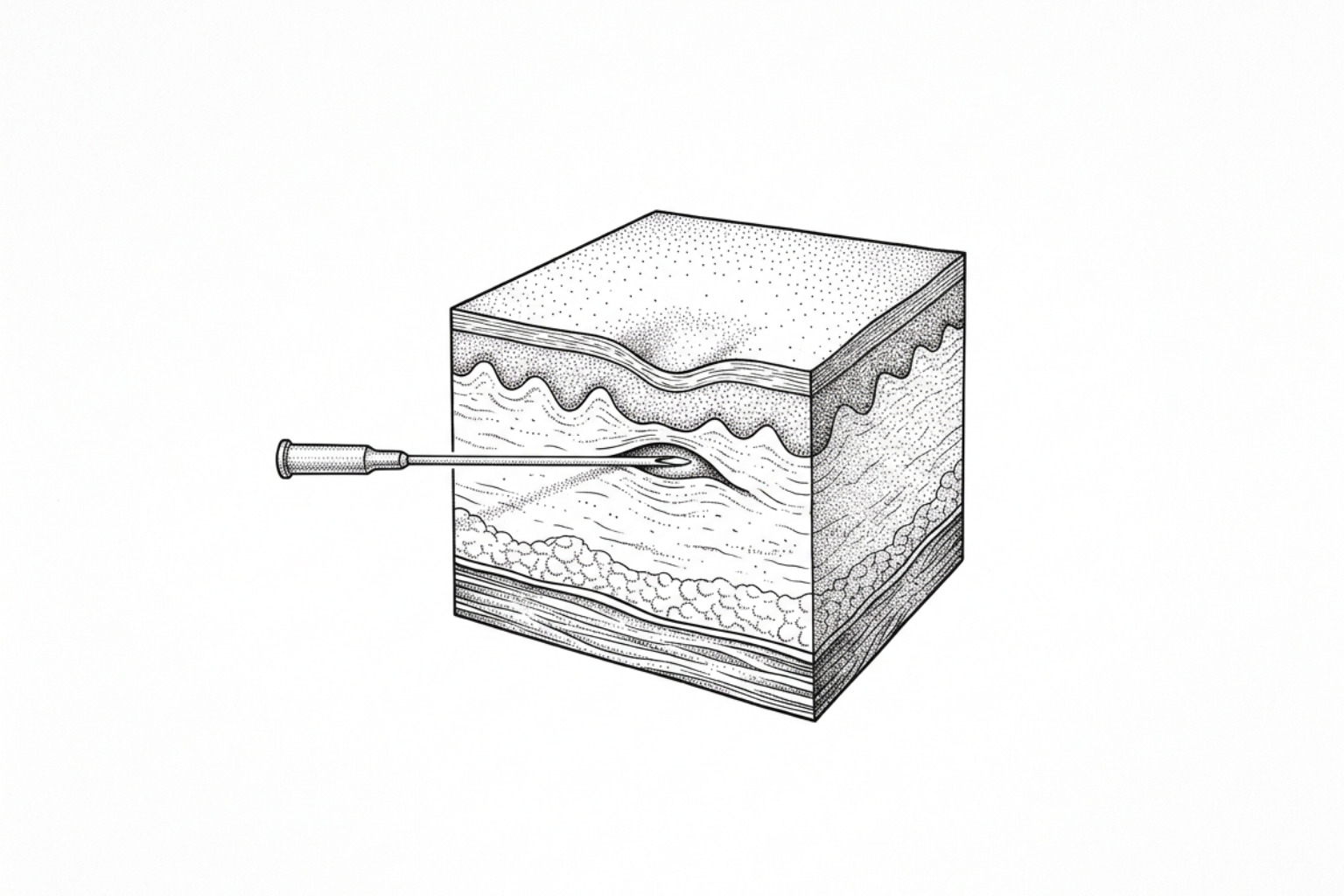
Subcision cuts the tethers pulling depressed acne scars downward, letting the skin rebound. Here's how the procedure works, which scars respond, the sharp-needle vs. blunt-cannula debate, and what recovery looks like.
What is subcision?
Subcision for scars is a minimally invasive surgical procedure used to treat depressed, tethered skin scars — most commonly those left behind by acne.
Here is a quick summary of what the evidence shows:
| Question | Answer |
|---|---|
| What does subcision treat? | Depressed (atrophic) scars, particularly rolling scars tethered by fibrous bands |
| How does it work? | A needle or cannula is inserted beneath the scar to break fibrous strands and stimulate collagen production |
| Best scar types | Rolling scars; tethered boxcar scars |
| Not suitable for | Ice-pick scars, keloid scars, raised hypertrophic scars |
| Sessions typically needed | 2–3 on average; moderate scarring may require up to 6 |
| Improvement range | 10–50% per session; higher with combination treatments |
| Downtime | 1–2 days for most patients |
| Results timeline | Visible at 2–4 months; remodeling continues for up to a year |
Acne scars affect up to 80–90% of adolescents who develop acne vulgaris, and the majority of those scars are atrophic — meaning the skin sits below the surrounding surface rather than rising above it. For many people, these depressions persist for years and prove resistant to topical treatments or surface-level procedures.
Subcision addresses a root cause that most surface treatments cannot reach: the fibrous strands anchoring the base of the scar to deeper tissue. First described by Orentreich and Orentreich in 1995 under the term subcutaneous incisionless surgery, the technique uses a needle or blunt cannula inserted through a small puncture in the skin to mechanically sever these tethers — allowing the depressed scar to lift and triggering the body's natural wound-healing response to lay down new collagen.
The procedure is performed in an outpatient setting under local anaesthesia. It is not a single-treatment cure, and it is not appropriate for every scar type. But for the right candidate, clinical evidence suggests it can produce meaningful, lasting improvement — particularly when combined with complementary treatments.
This guide covers the full clinical picture: how subcision works at a tissue level, which scar morphologies respond best, what the different instruments and techniques involve, what the research says about outcomes, and how to understand the realistic risks and recovery involved.
This content is for informational purposes only and does not constitute medical advice. Consult a qualified healthcare professional for diagnosis and treatment.
Subcision for scars definitions:
How subcision works
To understand why clinical consensus considers subcision for scars to be a foundational treatment in scar revision, the micro-anatomy of an atrophic scar must first be examined. When severe acne or inflammatory skin lesions heal, the normal dermal architecture is disrupted. In its place, the body deposits dense, disorganized collagen fibers.
In many atrophic scars, these fibrotic strands do not merely occupy the dermis; they extend vertically downward, anchoring the superficial dermis to the deeper subcutaneous tissue or superficial musculoaponeurotic system (SMAS). These bands act like structural anchors, pulling the skin surface downward and creating visible depressions or "potholes" in the skin contour.
Subcision functions through two distinct physiological mechanisms:
- Mechanical Release (Adhesiolysis): The clinician inserts a specialized instrument—such as a tri-beveled needle, blunt-tipped cannula, or surgical blade—into the subcutaneous plane parallel to the skin surface. By moving the instrument in a fan-like motion, the operator physically transects and severs these fibrous bands. This immediate mechanical release allows the tethered dermis to spring upward, restoring a more level skin contour.
- Controlled Microtrauma and Wound Healing: The physical movement of the instrument creates a controlled zone of microtrauma in the sub-dermal space. This trauma initiates the classic wound-healing cascade:
- Hemostasis: A small, localized hematoma (blood pool) intentionally forms in the newly created space beneath the released scar.
- Inflammation: Platelets in the hematoma release growth factors, recruiting fibroblasts to the site.
- Proliferation: Fibroblasts synthesize new extracellular matrix components, primarily Type III collagen, which is gradually replaced by stronger Type I collagen.
- Remodeling: Over the course of several months, this newly synthesized collagen reorganizes, acting as a natural structural scaffold that props up the scar from beneath and prevents the tissue from re-tethering.
Clinical guidance, such as the DermNet Subcision Guide, highlights that subcision's primary goal is this dual action of mechanical release and tissue remodeling. Without the mechanical release of these deep tethers, surface-level treatments like standard chemical peels or superficial microneedling cannot lift the depression. For a deeper understanding of how these mechanisms compare to other dermal lift methods, read the analysis of atrophic scar filling treatments.
Which scars subcision treats best
In clinical evaluations, patient selection is the single most critical factor determining the success of subcision for scars. Not all acne scars are structurally identical, and applying subcision to the wrong scar morphology will yield disappointing results.
To determine clinical suitability, dermatologists rely on qualitative classification systems, most notably the Goodman and Baron scar grading system. This system categorizes scars based on visibility and distensibility:
- Grade 1 (Macular): Flat, pigmented, or red marks that do not present contour changes.
- Grade 2 (Mild): Mild atrophic or hypertrophic scars that are easily covered by makeup or light shadows.
- Grade 3 (Moderate): Moderate atrophic or hypertrophic scars that are clearly visible at conversational distances but can be temporarily flattened by manually stretching the skin (distensible).
- Grade 4 (Severe): Severe atrophic or hypertrophic scars that are highly visible and cannot be flattened by manually stretching the skin (non-distensible).
A simple clinical diagnostic tool utilized during consultations is the skin stretch test. By placing fingers on either side of a depressed scar and gently pulling the skin taut, a clinician can assess its mechanical properties:
- If the scar flattens out completely, it is distensible and tethered by underlying fibrous bands. This indicates a high likelihood of response to subcision.
- If the scar remains depressed despite stretching, it is non-distensible. This suggests that the depression is caused by a structural loss of dermal tissue (volume deficit) or superficial epidermal tethering rather than deep fibrotic bands, requiring alternative or combinatorial treatments.
Rolling Scars: Where Subcision Excels
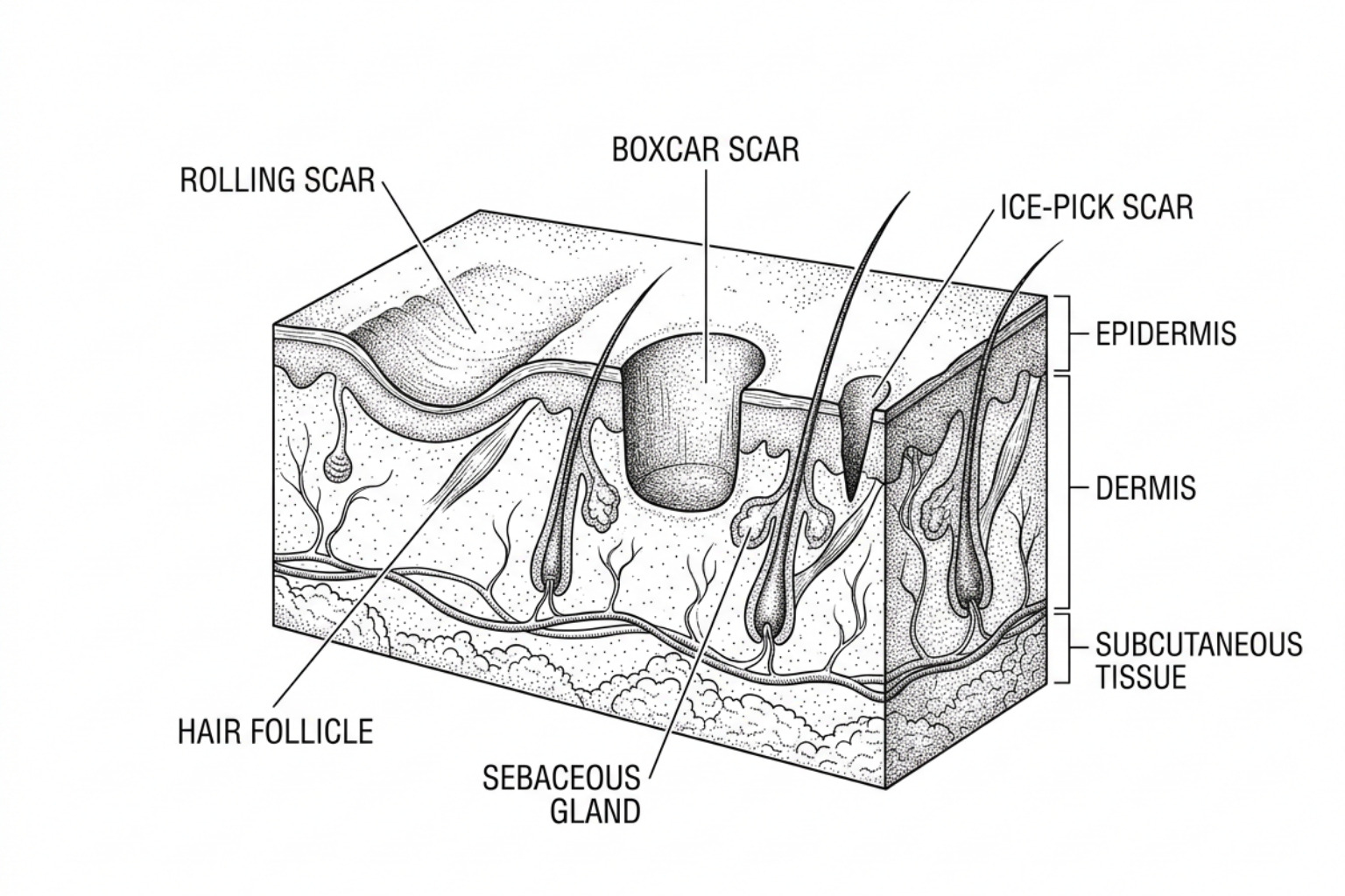
Rolling scars are the premier indication for subcision. These scars are typically wide (often greater than 4 mm in diameter), have gentle, sloping edges, and give the skin a wavy, uneven, or undulating appearance.
Because rolling scars are directly caused by fibrous anchoring at the dermal-subcutaneous junction, they are highly distensible. When performing subcision on rolling scars, the mechanical release of the tethers is highly effective.
Data shows that the degree of improvement from a single session of subcision typically ranges between 10% and 50%. However, achieving optimal outcomes usually requires a structured treatment program. An average patient requires between 2 to 3 subcision sessions, and those with more severe, deep-set rolling scars may require 3 to 6 visits spaced at least one month apart to allow for progressive collagen deposition.
To see real-world clinical timelines, you can read about the expected outcomes of subcision after one session or browse the clinical archives under the Tag: Acne Scars.
When subcision won't work: ice-pick and boxcar scars
In contrast, ice-pick and deep boxcar scars respond poorly to subcision as a standalone therapy:
- Ice-Pick Scars: These are narrow (less than 2 mm), deep, sharply demarcated tracts that extend vertically into the deep dermis or subcutaneous fat. Their primary pathology is a localized, complete loss of dermal tissue, not fibrous tethering. Standard subcision will not lift these scars because there are no horizontal or vertical bands to sever that would yield a significant lifting effect.
- Boxcar Scars: These are round or oval depressions with sharp, vertical, well-defined borders. While shallow boxcar scars may have some mild tethering, deep boxcar scars are characterized by structural tissue loss in the dermis.
Applying subcision to non-tethered scars can cause localized trauma, bruising, and swelling without producing any visible long-term lifting. For ice-pick and deep boxcar scars, alternative clinical modalities are required:
- TCA CROSS (Chemical Reconstruction of Skin Scars): High-strength trichloroacetic acid is precisely applied to the base of ice-pick scars to induce focal necrosis, triggering rapid localized collagen synthesis to close the tract.
- Punch Excision or Punch Elevation: The scar tissue is surgically punched out using a miniature biopsy tool, and the wound is either sutured closed or elevated to the level of the surrounding skin.
- Dermal Fillers: Temporary or semi-permanent fillers can be precisely injected to restore volume. For a comprehensive review of these volume-imparting options, explore the guides on dermal fillers for ice pick scars and ice pick scar fillers.
The tools and techniques doctors use
Over the past three decades, the surgical toolkit for performing subcision has evolved from simple hypodermic needles to highly specialized, blunt-tipped instruments and surgical wire systems. Each tool is designed to target specific tissue planes and minimize complications.
| Instrument | Sharpness | Primary Action | Best Suited For | Pros | Cons |
|---|---|---|---|---|---|
| Nokor Needle | Sharp (Tri-beveled, scalpel-like tip) | Cutting (horizontal slicing) | Isolated, dense, focal rolling scars | Highly effective at cutting tough, dense fibrotic bands | High risk of bleeding, hematoma, and post-procedure nodules; high operator dependency |
| Blunt Cannula | Blunt (requires a pilot needle entry) | Tearing/Puncturing (blunt dissection) | Widespread rolling scars, delicate anatomical zones | Significantly lower bruising and hematoma rates; minimal risk of nerve/vessel damage | May struggle to sever very thick, dense fibrotic bands (sometimes slips past them) |
| Taylor Liberator | Semi-sharp (paddle-shaped, multi-toothed blade) | Slicing/Scraping (large-scale subcision) | Severe, extensive, confluent rolling scars | Rapid treatment of large areas; highly uniform release | Requires tumescent anesthesia; higher immediate swelling; requires specialized training |
| Surgical Wire (Wire Scalpel) | Sharp (wire loop) | Sawing (mechanical shearing) | Large, deep, isolated atrophic plaques | Complete, comprehensive release of entire scar beds | Requires multiple entry/exit points; higher risk of localized tissue trauma |
Sharp Needle vs. Blunt Cannula Methods
The clinical choice between a sharp needle (such as the Nokor needle) and a blunt cannula represents a major decision point in scar revision.
The Nokor needle features a specialized scalpel-like tip that cuts horizontally as it is fanned through the tissue. While highly effective at slicing through dense, calcified fibrous bands, its sharp edges present a higher risk of cutting blood vessels and nerves.
In a comparative clinical study evaluating subcision complications, researchers found that 28.6% of patients experienced significant bleeding and bruising when treated with the sharp Nokor needle, compared to only 7.1% of patients treated with a blunt blade instrument Complications of Subcision for Acne Scarring.
Conversely, the blunt cannula method utilizes a flexible, non-sharp metal tube (typically 18-gauge to 22-gauge) that is introduced through a small pilot puncture made by a standard needle. Because the tip is blunt, it cannot cut blood vessels or nerves; instead, it pushes them aside, minimizing internal bleeding and bruising.
A landmark clinical trial published in the Journal of Cosmetic Dermatology demonstrated that cannula subcision achieved an 88% patient satisfaction rate and reduced the average number of visible scars from 24.8 to 12.8 per patient Subcision in acne scarring: A review of clinical trials.
Furthermore, split-face studies have demonstrated that while the clinical efficacy of the two methods is comparable at 6 months, the blunt cannula method offers a significantly faster recovery, less post-procedure pain, and a lower risk of post-inflammatory hyperpigmentation (PIH). For a detailed breakdown of this technique, refer to my guide on blunt cannula subcision.
Advanced Instruments: The Taylor Liberator and Wire Scalpels
For patients presenting with extensive, confluent areas of rolling scars—often described as "all-over" cheek scarring—traditional needle or cannula subcision can be tedious and require dozens of puncture sites. In these cases, advanced instruments like the Taylor Liberator or wire scalpels are utilized.
The Taylor Liberator is a long, sturdy, surgical-grade instrument featuring a paddle-shaped tip with three blunt-edged teeth. Rather than treating individual scars one by one, the clinician performs large-scale subcision, sweeping the instrument across the entire sub-dermal plane of the cheek.
To perform this safely and comfortably, tumescent anesthesia is mandatory. This technique involves infiltrating a large volume (typically 20 to 60 mL per cheek) of a highly diluted local anesthetic (lidocaine) mixed with epinephrine and sodium bicarbonate. Tumescent infiltration serves three critical clinical purposes:
- Hydrodissection: The volume of fluid physically expands the subcutaneous plane, separating the target dermis from deeper facial nerves and major blood vessels.
- Vasoconstriction: The epinephrine constricts blood vessels, dramatically reducing bleeding, bruising, and hematoma formation.
- Anesthesia: It provides complete, long-lasting pain relief across the entire treatment area.
Wire subcision (or wire scalpel subcision) represents another advanced mechanical approach. A fine surgical wire is threaded under the skin, entering at one point, wrapping around the margins of a large depressed scar area, and exiting through a second point. The clinician then performs a back-and-forth "sawing" motion with both ends of the wire, completely shearing the fibrous tethers across a broad plane.
According to a comprehensive review of surgical instruments in the Clinical, Cosmetic and Investigational Dermatology, wire subcision combined with immediate dermal support achieved some of the highest recorded clinical improvement scores for severe, bound-down scar plaques a comprehensive review of subcision instruments.
Combining subcision with other treatments
In clinical practice, subcision is rarely recommended as a standalone treatment. While subcision is highly effective at releasing fibrous bands, it does not address two other common components of acne scarring: superficial epidermal texture irregularities and localized volume deficits.
Furthermore, when subcision is performed alone, there is a risk of re-depression. As the internal wound heals, the newly forming scar tissue can contract, pulling the recently released dermis back down into its original depressed position.
To prevent re-depression and address all structural layers of the skin, clinicians utilize multimodal, combinatorial protocols:
- Subcision + Hyaluronic Acid (HA) Fillers: Immediately after severing the fibrous bands, the clinician injects a highly cohesive, cross-linked HA filler into the sub-dermal space. The filler acts as an immediate physical buffer, keeping the dermis elevated and preventing the severed tethers from re-attaching during early wound healing. Clinical studies show that subcision combined with HA filler achieved a 94.1% significant clinical improvement rate, compared to only 67.3% for subcision alone Subcision in acne scarring: A review of clinical trials.
- Subcision + Platelet-Rich Plasma (PRP) or Autologous Blood: Injecting PRP or the patient’s own blood (autologous blood subcision) into the sub-dermal space introduces concentrated growth factors that accelerate tissue regeneration, enhance collagen synthesis, and act as a temporary biostimulatory volume filler.
- Subcision + Collagen Biostimulators (PLLA / CaHA): For long-term structural support, biostimulators like Poly-L-lactic acid (PLLA) or Calcium Hydroxylapatite (CaHA) can be injected. These agents trigger a prolonged, low-grade inflammatory response that stimulates the body to produce its own natural Type I collagen over 6 to 12 months.
- Subcision + Energy-Based Devices (Fractional CO2 Laser / RF Microneedling): While subcision addresses the deep dermal and subcutaneous tethering, energy-based devices target the superficial dermis. Fractional CO2 lasers vaporize micro-columns of skin to trigger epidermal resurfacing, while Radiofrequency (RF) microneedling delivers thermal energy directly to the mid-dermis to tighten and smooth the skin texture.
- Subcision + TCA CROSS: In patients with mixed acne scarring (co-existing rolling and ice-pick scars), a combined approach is highly effective. The deep rolling scars are released via subcision, while the narrow ice-pick scars are treated in the same session using focal TCA CROSS.
A retrospective clinical study of 114 patients with severe, mixed acne scarring evaluated a combination protocol consisting of large-scale subcision (using a liberator blade), a superficial chemical peel, and fractional CO2 laser resurfacing performed in a single session. The researchers reported that 90% of patients were highly satisfied with their final results and did not require any further treatment sessions, demonstrating the power of synergistic, multi-layered protocols.
To explore how these surgical combinations compare to non-laser or needle-free alternatives, read the non-invasive acne scars complete guide. If you are dealing with raised or keloidal acne scars, subcision is contraindicated. Standard microneedling is contraindicated on keloid-prone skin as well: here is why.
Risks, side effects, and recovery
While subcision is a highly safe, outpatient procedure, it is still a form of subcutaneous surgery. Patients must have a realistic understanding of the expected side effects, potential complications, and recovery timeline.
What to expect afterward (downtime)
- Erythema (Redness) and Edema (Swelling): Swelling is immediate and typically peaks within 24 to 48 hours. This is actually beneficial in the short term, as the swelling temporarily plumps up the scars (a phenomenon known as the "swelling illusion"). Most swelling resolves within 3 to 7 days.
- Ecchymosis (Bruising): Bruising is common, particularly with needle-based subcision. It typically transitions from purple/blue to yellow and fades completely within 1 to 2 weeks.
What can go wrong
- Hematoma: An accumulation of blood under the skin. While a micro-hematoma is desired to stimulate healing, a large, expanding hematoma must be managed. Clinicians mitigate this by applying immediate, firm manual pressure for several minutes post-procedure. If a large hematoma does form, it may require clinical evacuation.
- Post-Inflammatory Hyperpigmentation (PIH): Temporary darkening of the skin at the needle entry points or across the treated area, most common in patients with darker skin tones (Fitzpatrick skin types IV–VI). Daily broad-spectrum SPF 50 sunscreen use and pre-treatment with tyrosinase inhibitors can minimize this risk.
- Subcutaneous Nodules (Lumps): These are firm, localized lumps that can form under the skin due to localized hematoma organization or excessive focal collagen production (fibroplasia). They occur in approximately 5–10% of cases, particularly when sharp needles are used in delicate areas like the temples or jawline. Most nodules resolve spontaneously within 2 to 3 months; persistent nodules can be treated with daily firm massage or low-dose intralesional corticosteroid/5-FU injections.
- Paresthesia (Numbness): Temporary nerve irritation or numbness caused by localized swelling or minor trauma to superficial sensory nerve branches. A review of clinical complications published in the Journal of Cosmetic Dermatology notes that subcision-induced paresthesia is almost always self-limiting and resolves spontaneously within 2 to 4 months as the nerves recover Complications of Subcision for Acne Scarring.
- Infection: Rare when proper aseptic techniques are maintained. Patients are advised to keep the entry points clean and avoid makeup for the first 24 hours.
- Hypertrophic Scarring: In rare instances (estimated at 5–10% of cases, primarily in patients with a genetic predisposition), the body's healing response can overcompensate, producing a raised scar. Subcision should be avoided in patients with a known history of keloid formation.
Aftercare for your skin
To optimize wound healing and minimize complications, patients are typically instructed to follow a strict post-care regimen:
- Cold Therapy: Apply cold compresses or ice packs wrapped in a clean cloth to the treated area for 10–15 minutes every hour on the day of the procedure to minimize swelling and bruising.
- Skin Barrier Support: Apply a thin layer of petroleum jelly or a healing ointment to the entry points twice daily until the skin has completely closed.
- Photoprotection: Avoid direct sun exposure and apply a broad-spectrum mineral sunscreen (zinc oxide-based, SPF 50+) daily to prevent post-inflammatory hyperpigmentation.
- Activity Restriction: Avoid vigorous exercise, heavy lifting, and saunas for the first 48 hours to minimize the risk of delayed bleeding or hematoma formation.
- Firm Massage: Starting on day 7 post-procedure, perform firm circular massage over any localized firm areas for 2–3 minutes twice daily to help smooth out developing collagen and prevent subcutaneous nodule formation.
For more detailed clinical guides on managing post-procedure healing and maximizing tissue regeneration, explore the Tag: Scar Treatment.
Key Takeaways: Is Subcision Right for You?
Subcision for scars remains one of the most mechanically sound and clinically proven treatments for depressed, tethered acne scars. By physically releasing the deep fibrous bands that pull the skin downward, it addresses a structural issue that surface-level lasers, chemical peels, and microneedling simply cannot reach.
However, because acne scarring is highly complex and often consists of multiple scar morphologies, subcision is rarely a standalone cure. Achieving optimal, smooth skin contouring requires a precise clinical evaluation, a customized treatment plan, and often a multimodal approach combining subcision with fillers, biostimulators, or energy-based devices.
Prior to beginning a scar revision journey, scheduling a comprehensive, professional clinical assessment is highly recommended to identify specific scar types and determine suitability for subcision.
To learn more about scar classification, explore interactive tools, or stay updated on the latest clinical research in wound healing:
- Get a professional, step-by-step evaluation with the Pillar Page: Scar Assessment.
- Browse the complete library of evidence-based guides on the Pillar Page: Topics.
- Stay informed on emerging dermatological therapies by visiting the Pillar Page: Subscribe.
Works Cited
- Barikbin, B., et al. "Blunt Blade Subcision: An Evolution in the Treatment of Atrophic Acne Scars." Dermatologic Surgery, 2017.
- Goodman, G. J., & Baron, J. A. "Postacne Scarring: A Qualitative Global Scarring Grading System." Dermatologic Surgery, 2006.
- Orentreich, D. S., & Orentreich, N. "Subcutaneous Incisionless (Subcision) Surgery for the Correction of Depressed Scars and Wrinkles." Dermatologic Surgery, 1995.
- "Subcision." DermNet NZ.
- Ahramiyanpour, N., et al. "Subcision in Acne Scarring: A Review of Clinical Trials." Journal of Cosmetic Dermatology, 2022.
- Sun, C., et al. "Complications of Subcision for Acne Scarring: Experience From Clinical Practice and Review of the Literature." Journal of Cosmetic Dermatology, 2024.
- Vempati, A., et al. "Subcision for Atrophic Acne Scarring: A Comprehensive Review of Surgical Instruments and Combinatorial Treatments." Clinical, Cosmetic and Investigational Dermatology, 2023.
This content is for informational purposes only and does not constitute medical advice. Consult a qualified healthcare professional for diagnosis and treatment.