Assess Scar Vascularity and Pliability Throughout the Post-Burn Maturation Phase
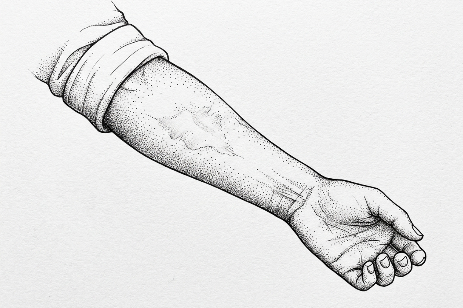
Burn scars keep changing long after the wound closes: redder and firmer for the first few months, peaking around six months, then slowly softening over 12 to 24 months. Here is what clinicians watch for and what helps.
How burn scars mature
Burn scars healing is a long remodeling process, not a single event. After a burn wound closes, clinicians track two important changes: vascularity (how red or blood-rich the scar appears) and pliability (how soft, flexible, or tight it feels). Research indicates that scars often become redder, thicker, and less flexible during the first few months, commonly peak around six months, and may continue to mature for 12 to 24 months.
Burn depth and healing time matter. Clinical guidance suggests burns that close within about 14 days are less likely to leave significant scars, while wounds that take three weeks or longer have a higher risk of hypertrophic scarring and contracture. Regular assessment can help identify a scar that is becoming increasingly raised, stiff, itchy, or movement-limiting, so a burn specialist or therapist can guide timely care.
This guide explains what changes are expected during post-burn scar maturation, how vascularity and pliability are assessed, and when conservative or clinical treatment may be appropriate. The Scar Healing Editorial Team provides this independent educational guide to support informed discussions with qualified healthcare professionals.
Discover more about burn scars healing:
How burn scars form
Understanding how scars form after thermal trauma requires looking at the cellular events in deep dermal tissue. A burn injury is not simply a mechanical cut; it triggers systemic and localized inflammatory cascades that disrupt the structural integrity of the extracellular matrix (ECM). According to comprehensive analyses in burn literature (Abarca-Pineda et al., 2025), when thermal injury extends into the reticular dermis, destruction of the basement membrane and microvasculature causes severe cellular dysregulation.
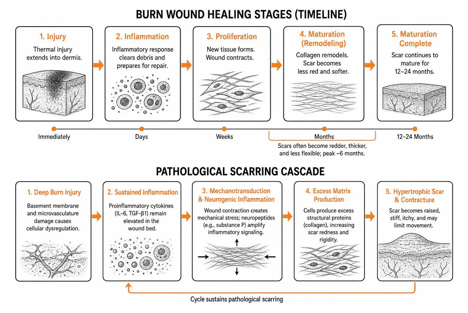
Mechanotransduction—the process by which cells convert physical mechanical tension into biochemical intracellular signals—plays a defining role in pathological scar development (Edger-Lacoursière et al., 2025). In uninjured skin, mechanical tension remains evenly distributed. However, post-burn wound contraction concentrates mechanical stress across the healing margin. This mechanical strain, coupled with neurogenic inflammation driven by neuropeptides such as substance P, stimulates local cell populations to produce excess structural proteins, increasing scar redness and rigidity.
Why scar cells overproduce collagen
During standard cutaneous wound repair, inflammation transitions smoothly into cell proliferation and eventual ECM remodeling. In deep burns, however, research on the molecular changes underlying hypertrophic scarring indicates that the inflammatory phase becomes sustained and dysregulated (Čoma et al., 2021). Elevated levels of proinflammatory cytokines, including interleukin-6 (IL-6) and transforming growth factor-beta 1 (TGF-β1), persist within the wound bed.
This persistent cytokine signaling drives the differentiation of resting dermal fibroblasts into contractile myofibroblasts, characterized by alpha-smooth muscle actin (α-SMA) expression. In normal physiological healing, myofibroblasts undergo programmed cell death (apoptosis) once the wound closes. In post-burn fibrosis, myofibroblasts evade apoptosis through mechanisms involving BCL-2 upregulation and matrix stiffness feedback loops (Faour et al., 2023). Consequently, these persistent cells produce excessive, disorganized type I and type III collagen bundles, leading to thick, non-compliant scar tissue.
Telling hypertrophic, keloid and contracture scars apart
Clinicians categorize burn-related scarring based on morphological characteristics, tissue behavior, and functional impact on the patient. Identifying the specific scar types: burn classification helps guide targeted clinical and conservative interventions.
- Hypertrophic Scars: These represent the most frequent complication following deep thermal injury, occurring in up to 70% to 90% of severe burn cases. They present as raised, firm, erythematous lesions that remain strictly within the original anatomical boundaries of the burn wound. Hypertrophic scars typically peak in vascularity and thickness within six months before entering a slow maturation phase.
- Keloid Scars: Characterized by uncontrolled proliferation, keloids extend beyond the original borders of the wound margins, invading surrounding healthy tissue. They demonstrate continuous collagen synthesis without a spontaneous regression phase and exhibit higher recurrence rates following surgical excision.
- Contracture Scars: When deep burns span mobile skin areas, flexor surfaces, or joints, the natural contractile forces exerted by myofibroblasts can physically shorten the skin envelope. Contractures lead to significant joint stiffness, functional impairment, and structural distortion of surrounding tissues.
| Scar Parameter | Hypertrophic Scar | Keloid Scar | Contracture Scar |
|---|---|---|---|
| Boundaries | Confined within original burn margins | Invades adjacent healthy tissue | Follows lines of skin tension/joints |
| Typical Onset | Within 1 to 3 months post-injury | Months to years after wound closure | Develops as scar matures and tightens |
| Natural Course | Peaks at ~6 months; slowly remodels | Persistent growth; rarely regresses | Progressive tightening without physical therapy |
| Primary Structural Feature | Disorganized, whorled collagen nodules | Thick, hyalinized collagen bundles | Shortened, unyielding fibrous bands |
| Functional Impact | Localized tightness, pruritus, dysesthesia | Cosmetic distortion, pain, hypersensitivity | Severe loss of joint range of motion (ROM) |
To track these structural variations objectively over time, burn care teams rely on validated clinical evaluation tools such as the Vancouver Scar Scale (VSS) and the Patient and Observer Scar Assessment Scale (POSAS). These clinical instruments assign numeric values to distinct parameters, including vascularity, pigmentation, pliability, and scar height, ensuring accurate documentation throughout recovery.
How burn scars heal over time
The timeline of burn scar maturation depends heavily on the initial depth of tissue destruction and how quickly the epithelial layer re-establishes itself. Superficial partial-thickness burns that complete re-epithelialization in less than 14 days carry a low risk of problematic scarring. Conversely, deep dermal wounds requiring three weeks or more to heal present a significantly elevated likelihood of developing pathological hypertrophic tissue (Abarca-Pineda et al., 2025). Proactive care, such as learning how to manage second-degree burns to minimize the risk of long-term scars, plays a critical role in mitigating these risks early on.
Redness over the first six months
During the initial six months post-closure, active tissue remodeling is characterized by robust neoangiogenesis. Immature scar tissue develops a dense, disorganized capillary network in response to local hypoxia and growth factor signaling. Clinically, this hypervascular state manifests as vivid erythema, ranging from bright pink to deep purple.
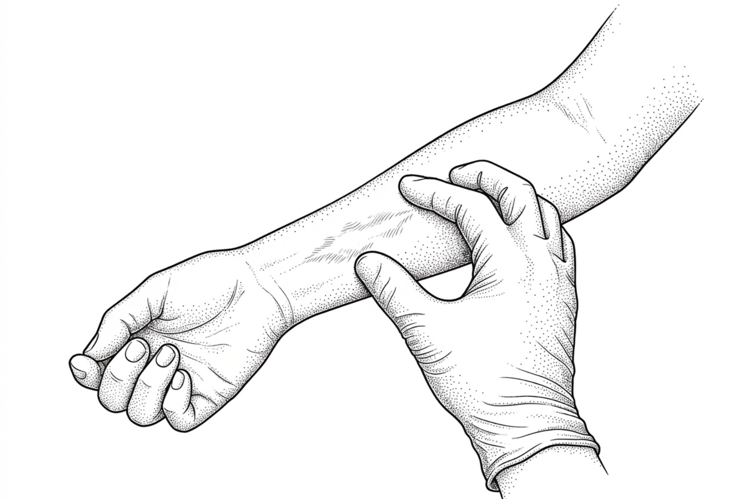
Clinicians monitor microvascular perfusion using tools like laser Doppler imaging and non-invasive colorimetry alongside standard visual grading. During this dynamic early phase, scars often feel warm to the touch, and heightened metabolic activity frequently correlates with intense itching (pruritus) and hypersensitivity.
Softening from 12 to 24 months
As the scar approaches the 12- to 24-month window, cellular activity within the dermal matrix gradually shifts. Capillary density begins to normalize through natural vascular regression, leading to a visible fading of scar erythema from deep red to pale pink or flesh-toned hues.
Concurrently, endogenous matrix metalloproteinases (MMPs) slowly break down and realign chaotic collagen bundles into more organized, parallel configurations. This structural reorganization improves scar pliability, reducing firmness and skin tension. However, because dense scar tissue lacks functional elastin fibers, full pre-injury elasticity is rarely recovered, emphasizing the importance of long-term scar surveillance and therapy.
Non-surgical treatment options
Non-invasive therapies form the initial foundation of post-burn rehabilitation. Evidence indicates that early conservative physical modalities help reduce microvascular congestion, relieve physical tension across tissue planes, and regulate aberrant fibroblast activity (Edger-Lacoursière et al., 2025). For a broader exploration of non-operative approaches, see the non-surgical burn scars complete guide.
Pressure garments and silicone sheets
First-line clinical guidelines frequently recommend combining custom pressure garment therapy with medical-grade silicone treatment.
- Pressure Garment Therapy: Custom-tailored elastic garments apply continuous, graduated pressure (typically 15 to 30 mmHg) over healing areas. This mechanical compression reduces local capillary blood flow, induces mild tissue hypoxia to downregulate fibroblast proliferation, and mechanically encourages newly deposited collagen fibers to align parallel to the epidermal plane. For optimal efficacy, clinical protocols recommend wearing garments for up to 23 hours per day throughout the active maturation phase.
- Topical Silicone Gels and Sheeting: Silicone acts primarily by restoring epidermal barrier function. By occluding the stratum corneum, silicone sheets significantly reduce transepidermal water loss (TEWL). This deep hydration normalizes keratinocyte signaling, which downstream decreases the release of profibrotic cytokines that stimulate myofibroblasts. Silicone sheeting is generally applied for 12 to 24 hours daily once the skin has completely re-epithelialized.
Hydration, Gentle Massage, and Sun Protection
Burn injuries permanently damage or destroy dermal appendages, including sebaceous and sweat glands. This loss compromises the skin's natural lipid barrier, leaving newly healed tissue prone to severe xerosis, cracking, and breakdown. Applying bland, fragrance-free emollients several times daily helps restore moisture balance. Understanding how to protect healing skin from secondary environmental insults is essential for long-term recovery.
- Targeted Scar Massage: Gentle, circular manual massage with hydrating emollients aids in desensitizing hypersensitive cutaneous nerves, mobilizes restricted tissue planes, and temporarily alleviates pruritus. However, clinicians caution against overly forceful or aggressive friction massage during early healing stages, as excessive shear strain can trigger secondary inflammation and disrupt delicate anchoring fibrils (Edger-Lacoursière et al., 2025).
- Sun Protection: Immature scar tissue contains unstable melanocyte populations and compromised barrier defenses, making it susceptible to UV-induced damage. Unprotected exposure to sunlight can trigger prolonged post-inflammatory hyperpigmentation that may become permanent. Patients should apply broad-spectrum sunscreen with SPF 50+ regularly and wear UV-protective clothing over healed areas for at least 12 to 18 months post-injury.
Laser and surgical options
When conservative modalities prove insufficient to manage severe thickness, joint restrictions, or persistent redness, advanced clinical interventions may be incorporated. Comprehensive burn care protocols balance conservative and operative therapies based on scar maturity and functional limitations (Abarca-Pineda et al., 2025). A detailed review of medical and procedural options is available in the burn scars treatment guide.
Using laser for burn scars
Laser technology has become a cornerstone of modern burn scar management, offering precise tissue remodeling with minimal recovery downtime (Ma et al., 2023). Different laser platforms target distinct physical components of the scar matrix:
- Pulsed Dye Laser (PDL, 585–595 nm): PDL selectively targets intravascular hemoglobin via selective photothermolysis. By coagulating excess microvessels within the scar, PDL reduces erythema, lowers metabolic activity, and improves overall POSAS scores. Systematic reviews indicate that PDL provides substantial improvements in scar vascularity and height (Ma et al., 2023).
- Fractional Ablative Carbon Dioxide (CO₂, 10,600 nm) Lasers: Fractional CO₂ devices create microscopic vertical columns of thermal injury (microscopic treatment zones) that extend deep into the fibrotic reticular dermis. This controlled thermal ablation vaporizes rigid collagen cords, triggers heat-shock proteins, and stimulates matrix metalloproteinases to remodel stiff scar tissue, improving overall pliability.
- Timing and Treatment Intervals: Clinical meta-analyses indicate that laser therapy performed at intervals of 4 to 8 weeks balances efficacy with adequate tissue recovery (Ma et al., 2023). While early vascular laser intervention can help manage active erythema, fractional ablative lasers are often applied to mature, dense fibrous bands to restore flexibility.
Surgical release and regenerative options
Severe burn contractures that restrict joint movement, compress neurovascular bundles, or cause structural distortions generally require formal surgical revision. The scar revision treatment complete guide outlines standard surgical pathways:
- Z-Plasty and Geometric Transposition Flaps: These techniques lengthen contracted scar bands and redirect the vectors of mechanical skin tension away from primary lines of joint motion.
- Excision and Grafting: Dense, unyielding scar tissue is surgically excised and replaced with split-thickness skin grafts (STSG), full-thickness skin grafts (FTSG), or local tissue flaps to restore soft-tissue coverage.
- Autologous Fat Grafting (Lipofilling): Injecting purified adipose tissue beneath fibrotic scars introduces adipose-derived stem cells (ADSCs). Early clinical observations suggest these cells secrete regenerative paracrine factors that soften rigid scar beds, improve dermal elasticity, and reduce neuropathic pain (Abarca-Pineda et al., 2025).
Preventing contractures and supporting recovery
Recovering from a severe burn injury involves functional physical rehabilitation alongside cosmetic management. Because mature scars undergo progressive mechanical contraction, systematic physical and occupational therapy must begin immediately after acute wound closure.
Without consistent active motion and positioning regimens, joint contractures can develop rapidly. Clinical literature indicates that up to 20% to 30% of surgical contracture releases experience recurrence if postoperative rehabilitation and splinting protocols are suboptimal (Edger-Lacoursière et al., 2025). Multi-angle splinting, serial casting, and progressive active range-of-motion (AROM) exercises provide sustained mechanical elongation that counteracts myofibroblast shortening.
Relieving pain and itching
Post-burn pruritus and neuropathic dysesthesia are among the most persistent symptoms reported by survivors:
- Mechanisms: Regenerating cutaneous nerve endings within the remodeling dermis display heightened mechanical and thermal sensitivity. In addition, resident mast cells periodically degranulate, releasing histamine, substance P, and inflammatory mediators directly into the healing matrix.
- Management Strategies: First-line clinical interventions combine oral second-generation H1-antihistamines with topical agents containing pramoxine, camphor, or menthol to cool peripheral receptors. Regular application of deeply hydrating, fragrance-free emollients helps maintain skin hydration, reducing the urge to scratch and preventing secondary excoriation.
Emotional recovery after a burn
The physical changes caused by burn scars frequently alter body image, leading to heightened anxiety, social withdrawal, or post-traumatic stress. Holistic rehabilitation models emphasize that psychological recovery is as vital as physical wound healing.
Trauma-informed counseling, cognitive behavioral therapy (CBT), and peer support networks help survivors process body image changes, build practical coping strategies, and regain confidence during social reintegration.
Frequently Asked Questions About Burn Scar Maturation
How long does it take for a burn scar to fully mature?
Most burn scars require between 12 and 24 months to complete their biological maturation. During this timeframe, microvascular blood flow slowly declines, leading to reduced redness, while internal collagen remodeling gradually softens and flattens the tissue. The precise duration varies based on the initial depth of the burn, patient age, genetic factors, and adherence to conservative therapies.
What are the early signs that a burn is forming a hypertrophic scar?
Key clinical indicators include a wound that requires longer than two to three weeks to achieve full closure, persistent and intensifying redness beyond the first month, noticeable thickening or hardening of the tissue, and severe, escalating itching (pruritus).
When is the ideal time to start conservative scar management?
Conservative modalities should begin as soon as the wound bed achieves complete re-epithelialization and the newly formed skin is structurally stable—typically within days of final closure. Therapeutic hydration, custom pressure garments, and silicone sheeting are most effective when introduced early in the post-closure period, as discussed in the clinical guide on when to start scar treatment after surgery.
Conclusion
Managing post-burn scar maturation requires ongoing tracking of scar vascularity and pliability. While the biological remodeling phase extends over one to two years, consistent application of multimodal conservative care—including pressure garments, silicone therapy, targeted hydration, and specialized laser interventions—helps guide tissue organization. By monitoring physical changes and collaborating closely with a multidisciplinary burn care team, patients can optimize both functional mobility and long-term aesthetic outcomes.
Take an active step in your recovery journey: start your scar assessment today to understand your scar's maturation stage and explore evidence-based care options.
This content is for informational purposes only and does not constitute medical advice. Consult a qualified healthcare professional for diagnosis and treatment.
Works Cited
- Abarca-Pineda YA, Alaniz IC, Ibraheam A, et al. “Conventional and Emerging Surgical and Non-surgical Approaches to the Management of Skin Burn Scars: A Comprehensive Review.” Cureus, 2025.
- Edger-Lacoursière Z, Zhu M, Jean S, Marois-Pagé E, Nedelec B. “Evidence Supporting Conservative Scar Management Interventions Following Burn Injury: A Review Article.” Journal of Burn Care & Research, 2025.
- Ma Y, Barnes SP, Chen YY, Moiemen NS, Lord JM, Sardeli AV. “Influence of scar age, laser type and laser treatment intervals on adult burn scars: A systematic review and meta-analysis.” PLoS One, 2023.
- Čoma M, Fröhlichová L, Urban L, et al. “Molecular Changes Underlying Hypertrophic Scarring Following Burns Involve Specific Deregulations at All Wound Healing Stages (Inflammation, Proliferation and Maturation).” International Journal of Molecular Sciences, 2021.
- Faour S, Farahat M, Aijaz A, Jeschke MG. “Fibrosis in burns: an overview of mechanisms and therapies.” American Journal of Physiology-Cell Physiology, 2023.