A Practical Guide to Improving Collagen Remodeling in Scars
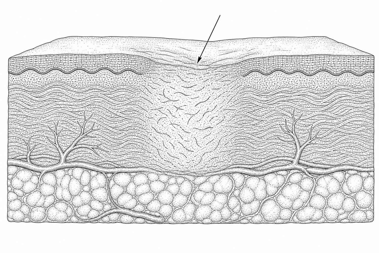
Scar collagen keeps remodeling for months. Here's how lasers, microneedling, silicone, and simple home care can steer it toward smoother, flatter skin.
How collagen reshapes a scar over time
Understanding how to improve collagen remodeling in scars is one of the most clinically relevant questions in dermatology — and one of the most frequently misunderstood by patients managing persistent acne marks, surgical scars, or injury-related skin changes.
Here is a direct answer to that question, based on current evidence:
Evidence-based strategies to improve collagen remodeling in scars:
- Silicone gel or silicone sheet therapy — applied daily once the wound has fully closed; evidence suggests it regulates hydration and reduces excessive collagen deposition
- Fractional laser therapy — stimulates orderly collagen synthesis by creating controlled micro-injuries in the dermal layer
- Microneedling — induces a controlled wound healing response at depth, prompting fibroblasts to produce new, better-organized collagen
- Pressure therapy — compression garments exerting 20–30 mmHg reduce mechanical tension and flatten raised scars
- Scar massage — initiated 2–3 weeks post-injury, cross-friction and circular techniques help break down disorganized fibrous tissue
- Corticosteroid injections — suppress overactive fibroblasts in hypertrophic and keloid scars
- Nutritional support — adequate Vitamin C, zinc, and protein intake supports healthy collagen synthesis
- Sun protection (SPF 30+) — prevents UV-induced disruption of the remodeling process and permanent discoloration
Scars form because the body prioritizes speed over perfection when closing a wound. The result is collagen that is structurally different from normal skin — often disorganized, mechanically weaker, and resistant to natural repair. Collagen makes up roughly three-quarters of the dry weight of human skin, and when its structure is disrupted by injury, the remodeling process that follows can last up to a full year. How well that process goes determines whether a scar remains raised, sunken, or gradually fades toward the skin's surface.
The challenge is that collagen remodeling is not passive. It is driven by a precise sequence of cellular signals, immune responses, and mechanical forces — all of which can be influenced, for better or worse, by the treatments applied and the choices made during recovery.
The Scar Healing Editorial Team has reviewed the latest peer-reviewed research across dermatology, wound biology, and regenerative medicine to compile what follows — a practical, evidence-grounded guide to the most effective strategies currently available.
The stages of wound healing
To understand how to alter a scar's final appearance, it is essential to first examine the biological environment in which it forms. Cutaneous wound healing is a highly coordinated, dynamic progression divided into four overlapping phases: hemostasis, inflammation, proliferation, and maturation (or remodeling).
During the inflammatory phase (which typically persists for 3 to 5 days), immune cells clear debris and release pro-inflammatory cytokines. The proliferative phase (spanning roughly 3 to 4 weeks) is characterized by angiogenesis and the rapid migration of fibroblasts. These cells synthesize a temporary extracellular matrix (ECM) to close the gap.
In clinical analyses of wound dynamics, the transition from late proliferation to early maturation is where the trajectory of a scar is decided. Pathological scarring occurs when these phases are disrupted — particularly by prolonged inflammation or sustained mechanical tension. Under these stresses, standard fibroblasts differentiate into highly contractile myofibroblasts, which overproduce matrix proteins and lock the tissue into a rigid, elevated state. For a detailed breakdown of this entire physiological sequence, read the comprehensive guide on How Scars Form.
How Scars Form: The Role of Collagen Types I and III
In healthy, uninjured adult skin, the dermal matrix is dominated by Type I collagen, which provides structural strength, alongside a smaller proportion of Type III collagen, which offers elasticity. Mechanically, normal skin maintains a Type I to Type III collagen ratio of approximately 4:1 (quantified in some control studies as 3.2 ± 0.5).
During the early proliferative phase of healing, the body rapidly deposits Type III collagen because its smaller, highly flexible fibrils can be synthesized quickly to restore structural integrity. This temporarily shifts the Type I to Type III ratio down to approximately 2:1 (or 1.8 ± 0.2 in untreated immature scars).
During the subsequent remodeling phase, the body produces matrix metalloproteinases (MMPs) to degrade this temporary Type III collagen and replace it with sturdier Type I collagen fibers. However, even under optimal conditions, a fully matured scar never recovers the original basket-weave architecture of healthy skin; its parallel, densely packed collagen bundles mean that a healed scar typically achieves a maximum of only 70% to 80% of the tensile strength of uninjured tissue. To understand the cellular machinery behind this protein production, explore the resource on Protein Synthesis Wound Healing.
What controls collagen-making cells
The synthesis, deposition, and structural organization of collagen are governed by a network of signaling molecules. Chief among these are the transforming growth factor-beta (TGF-β) isoforms, which exert contrasting effects on the healing microenvironment:
- TGF-β1 and TGF-β2: These are highly pro-fibrotic cytokines. They drive the differentiation of fibroblasts into alpha-smooth muscle actin (α-SMA)-expressing myofibroblasts, stimulate excessive collagen deposition, and suppress matrix-degrading enzymes.
- TGF-β3: In contrast, TGF-β3 limits the inflammatory reaction, promotes orderly collagen fiber alignment, and is highly expressed in scarless fetal wound healing.
Other key cytokines include interleukin-6 (IL-6), a pro-inflammatory mediator that can perpetuate fibrotic loops, and interleukin-10 (IL-10), an anti-inflammatory cytokine that downregulates collagen synthesis to encourage an embryonic-like, scar-free healing environment. In reviews of the biochemical literature, targeting this cytokine balance remains a primary focus of advanced scar therapies. Researchers seeking a deeper molecular analysis can consult the Scientific research on cytokine modulation of fibroblast collagen production.
The main types of scars
When the regulatory mechanisms of the remodeling phase fail, the result is pathological scarring. Clinically, these are categorized into three primary structural types: hypertrophic scars, keloids, and atrophic scars.
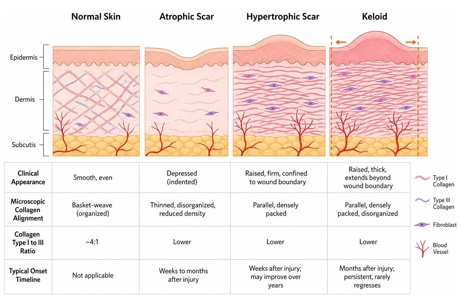
Hypertrophic scars are raised, red, and rigid, but they remain strictly confined within the boundaries of the original wound and frequently undergo slow regression over several years. Keloids, however, behave like benign collagenous tumors; they grow invasively beyond the margins of the original injury, rarely regress, and carry a high rate of recurrence after surgical excision. Atrophic scars present as depressions or indentations, resulting from a localized deficit of collagen support during the healing of inflammatory conditions like acne or varicella.
To determine the exact nature and severity of a scar before initiating treatment, practitioners rely on standardized clinical scales. These diagnostic parameters are detailed in the guide on Scar Assessment.
| Scar Type | Clinical Appearance | Microscopic Collagen Alignment | Collagen Type I to III Ratio | Typical Onset Timeline |
|---|---|---|---|---|
| Normal Mature Scar | Flat, pale, soft | Parallel to skin surface | ~4:1 | 12 to 24 months |
| Hypertrophic Scar | Raised, red, confined to wound | Fine, wavy, parallel bundles | ~6:1 (elevated Type I) | 1 to 2 months post-injury |
| Keloid | Raised, bulbous, spreads beyond wound | Random, thick, disorganized | ~17:1 (extreme Type I dominance) | Months to years post-injury |
| Atrophic Scar | Sunken, pitted, thin | Scant, fragmented network | Variable (overall collagen deficit) | Weeks to months post-injury |
How collagen differs in scars
Under polarized light microscopy, the structural differences between these scars become clear. In hypertrophic scars, the collagen fibers are arranged in wavy, parallel patterns relative to the epidermis. In keloids, the fibers form random, thick, highly disorganized bundles.
This disorganization is driven in part by Lysyl Oxidase (LOX), an enzyme responsible for the covalent cross-linking of collagen molecules to stabilize fibrillar structures. In keloids, LOX activity is approximately 3 times higher than in normal skin, resulting in excessive cross-linking, extreme tissue stiffness, and resistance to natural enzymatic degradation.
Furthermore, while the average collagen fibril diameter in normal skin ranges from 110 to 130 nm, it is significantly reduced in keloids (~76 nm) and hypertrophic scars (~60 nm). In evaluations of this paradox — thinner individual fibrils forming much thicker, stiffer macroscopic bundles — evidence suggests that rapid, hyperactive procollagen synthesis leads to incomplete cleavage of N-terminal propeptides. The remaining propeptides cause steric hindrance, preventing individual fibrils from growing to their normal lateral dimensions while packing tightly into rigid, non-functional masses.
Treatments that rebuild collagen
When managing mature or highly symptomatic scars, home care alone is often insufficient. Clinical, evidence-based interventions are required to physically disrupt pathological matrix configurations and restart cellular remodeling. For an overview of which clinical procedures perform best across various scar types, refer to the guide on What Works for Scars.
How laser therapy works
Energy-based devices, particularly the fractional CO2 laser (operating at a wavelength of 10,600 nm), have altered how we approach scar revision. Rather than ablating the entire skin surface, fractional photothermolysis creates thousands of microscopic treatment zones (MTZs) of controlled thermal damage while leaving the surrounding tissue intact.
This triggers a highly controlled, biphasic cellular response:
- Immediate Phase (Days 0–10): The thermal shock downregulates Type I and Type III procollagen mRNA expression, preventing immediate fibrotic rebound. Simultaneously, it upregulates key matrix metalloproteinases (MMPs), which initiate the degradation of old, rigid collagen.
- Delayed Phase (Day 10 to 3 months+): The laser induces a phenotypic switch in macrophages from pro-inflammatory to pro-reparative (Arg1-positive) states. It downregulates α-SMA expression in hyperactive myofibroblasts, relieving tissue contracture, and prompts the orderly deposition of new, parallel collagen fibrils.
In analyses of clinical trials, this dual action of removing pathological collagen while stimulating fresh matrix synthesis makes fractional CO2 lasers exceptionally effective for both hypertrophic and atrophic scars. For a deeper look at the cellular pathways involved, see the Scientific research on fractional CO2 laser mechanisms or read the clinical overview of CO2 Laser Scar Removal.
How microneedling works
For patients seeking a non-thermal alternative, microneedling (or collagen induction therapy) is a highly reliable clinical option. This procedure uses medical-grade, sterile needles to puncture the dermis, creating thousands of physical micro-channels.
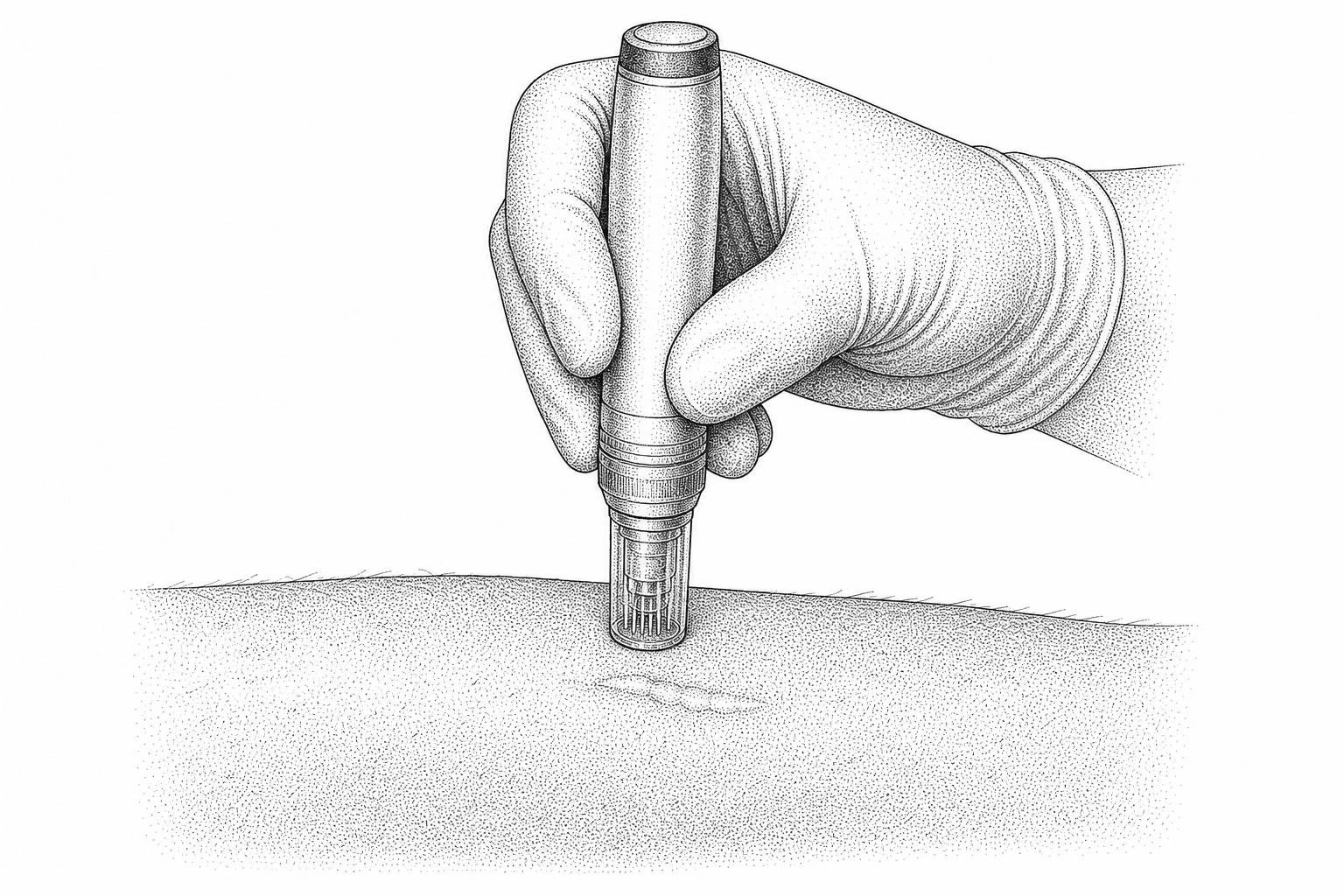
These micro-injuries trigger a localized healing cascade without damaging the overlying epidermis. The physical disruption of the old collagen scaffolding prompts platelets and fibroblasts to release growth factors, such as basic fibroblast growth factor (bFGF) and TGF-β3.
Clinical parameters are vital for safety and efficacy:
- Needle Depth: Typically ranges from 0.5 mm to 2.5 mm, dynamically adjusted based on the scar's thickness and location.
- Frequency: Most patients require a series of 3 to 6 sessions spaced 4 to 6 weeks apart.
- Efficacy: Data shows that most patients experience a 50% to 80% improvement in scar texture after completing a full clinical series.
To explore how this mechanical stimulation compares to other resurfacing tools, consult the guide on Microneedling Benefits Scars.
Silicone sheets and tension relief
Silicone gel and silicone sheets remain the international gold standard for non-invasive scar management. The primary mechanism of silicone is not chemical, but physical: it mimics the barrier function of the stratum corneum, reducing transepidermal water loss (TEWL).
This localized hydration signals the overlying keratinocytes to produce fewer cytokines that stimulate fibroblasts. This downregulates IL-1 expression and limits excessive collagen deposition.
Additionally, mechanical tension on a healing wound activates mechanotransduction pathways (such as YAP/TAZ and FAK) that promote myofibroblast activity. Silicone sheets, especially when combined with pressure therapy (exerting 20 to 30 mmHg of pressure), help shield the healing tissue from these lateral stretching forces, encouraging a flatter, more pliable scar. To understand how to incorporate this into a recovery protocol, see the clinical review of Hypertrophic Scar Silicone Gel.
New and experimental treatments
The field of scar revision is shifting toward molecular and cellular therapies designed to guide tissue regeneration rather than simply treating existing scar tissue.
Experimental nanoparticle treatments
One of the most promising areas of research involves nanodrug delivery systems (NDDSs) and advanced biomaterial scaffolds. Traditional topical creams struggle with poor transdermal penetration, rarely reaching the deep dermis where active myofibroblasts reside.
To overcome this, researchers are developing stimuli-responsive nanocarriers that can penetrate the skin barrier and release active compounds in response to microenvironmental cues, such as localized oxidative stress or acidic pH.
Recent laboratory and animal studies have highlighted several innovative platforms:
- Graphene-Collagen Cryogels: Highly porous scaffolds that physically modulate the mechanical environment of the wound while serving as delivery vehicles.
- Stem Cell-Derived Apoptotic Vesicles (ASCs-ApoVs): These vesicles carry biological signaling factors that suppress myofibroblast proliferation, downregulate α-SMA, and promote M2 macrophage polarization.
- Stromal Vascular Fraction (SVF) Gel: An autologous fat derivative rich in regenerative cells and extracellular matrix components, which has shown synergistic effects when combined with fractional CO2 lasers to treat depressed scars.
To review the primary literature on these technologies, you can read the Scientific research on fibroblast-targeted nanodelivery systems, the Scientific research on graphene-collagen cryogels, or the clinical trial analyzing Scientific research on fractional CO2 combined with SVF gel.
Drugs that block scarring
Another emerging paradigm is the repurposing of systemic medications into targeted topical therapies to block the molecular pathways of fibrosis:
- Pirfenidone: An antifibrotic agent that inhibits fibroblast proliferation and reduces TGF-β expression. In pediatric burn trials, a topical 8% pirfenidone gel led to significant improvements in Vancouver Scar Scale (VSS) scores.
- Angiotensin II Blockade (Losartan & Enalapril): Commonly used cardiovascular medications, such as losartan (an ARB) and enalapril (an ACE inhibitor), have secondary biochemical pathways that suppress TGF-β1. Clinical studies show that topical 5% losartan ointment can reduce keloid height and vascularity.
- Adenosine A2A Receptor (A2AR) Antagonists: In animal models, blocking the A2A receptor (using antagonists like ZM241385) starting around two weeks post-injury prevented the excessive accumulation of Type III collagen, restored the natural 4:1 collagen ratio, and increased the tensile strength of the healed wound.
To evaluate the clinical evidence supporting these pharmacological approaches, consult the Scientific research on medications for scar minimization.
What you can do at home
While clinical treatments are powerful, everyday mechanical and metabolic support is essential for optimal dermal reconstruction. To protect the skin's barrier during daily activities, refer to the practical guide on How Protect Healing Skin.
Scar massage
Scar massage is a highly effective, low-cost mechanical therapy when performed consistently. Clinical guidelines recommend waiting until the wound has completely closed with no active scabbing or remaining sutures (typically 2 to 3 weeks post-injury or surgery) before beginning.
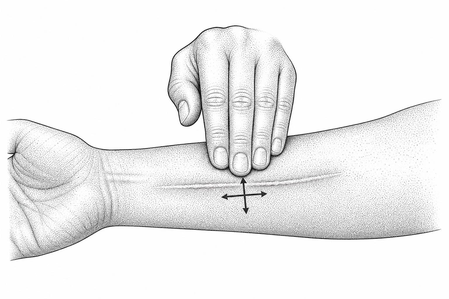
The physiological benefits of massage are rooted in mechanobiology. Applying physical pressure to the tissue:
- Disrupts the parallel, rigid cross-linking of pathological collagen fibers.
- Increases local blood flow, delivering essential oxygen and nutrients to the remodeling tissue.
- Stimulates localized matrix metalloproteinase (MMP) activity to help soften hard scar tissue.
Massage Protocol: Perform the massage for 5 to 10 minutes, 2 to 3 times daily. Use circular or cross-friction movements (massaging perpendicular to the direction of the scar) with enough pressure to blanch the skin slightly, but never to the point of pain. To avoid skin irritation, use a simple water-based lubricant or silicone gel. For a complete guide on these techniques, see the Scar Tissue Massage Guide 2026.
Nutrition and sun protection
The metabolic demands of collagen remodeling require specific micronutrients. Without these cofactors, fibroblast activity slows, resulting in weaker tissue. For a comprehensive review of these nutrients, read the Best Vitamins for Scar Healing Guide.
- Vitamin C (Ascorbic Acid): An essential cofactor for the enzymes prolyl hydroxylase and lysyl hydroxylase, which stabilize the collagen triple-helix structure.
- Zinc: A critical cofactor for matrix metalloproteinases (MMPs), which are required to degrade old, disorganized collagen.
- Protein (Amino Acids): L-arginine and L-glutamine support cellular proliferation and collagen synthesis.
- Sun Protection (SPF 30+): UV radiation triggers inflammatory cascades and upregulates melanocyte activity, which can lead to permanent hyperpigmentation and disrupt the delicate enzymatic balance of the remodeling phase. Dermatologists advise applying a broad-spectrum, mineral-based sunscreen daily for at least one year post-injury.
Frequently asked questions
Does collagen supplementation help repair scar tissue?
There is currently no strong clinical evidence showing that oral collagen supplements directly target or selectively remodel scar tissue. While oral collagen peptides are broken down into beneficial amino acids (such as proline and glycine) that support overall skin hydration and elasticity, they are distributed systemically. They cannot be directed specifically to a scar site. Consuming adequate dietary protein is generally sufficient to support the body's natural healing processes.
What is the timeline for scar remodeling to complete?
The maturation and remodeling phase is a slow process that begins around the second or third week post-injury and continues for up to 12 to 24 months. During the first 3 to 6 months, the scar is highly active, often presenting as red, raised, and firm. Over the course of a year, cellular activity gradually declines, blood vessels regress, and the scar flattens and fades. Interventions are most effective when initiated during this active, first-year window.
Can old, mature scars still undergo remodeling?
Yes, but mature scars require more active physical or clinical stimulation. Once a scar has matured (typically after one year), its collagen network is stable and cellular turnover is low. To restart the remodeling process, treatments like fractional CO2 lasers, microneedling, or surgical revision are used to create controlled micro-injuries, signaling the body to initiate a fresh, highly organized wound healing cascade.
Key takeaways
Improving collagen remodeling in scars requires a patient, multi-faceted approach. Because the remodeling phase is highly dynamic, successful scar management depends on matching the right treatment to the scar's specific developmental stage. Whether utilizing clinical interventions like fractional lasers and microneedling, or consistent at-home strategies like silicone therapy, scar massage, and strict sun protection, the goal remains the same: guiding the body away from rigid, disorganized fibrosis and toward flexible, orderly tissue regeneration.
Because skin type and genetic background heavily influence fibroblast activity, seeking a personalized clinical assessment is highly recommended to design a treatment plan tailored to the skin's unique needs. For individuals with deeper skin tones who are more prone to hyperpigmentation or keloid formation, specialized guidance is particularly important; specialized clinical insights are available in the Comprehensive Guide to Scar Removal for Dark Skin.
Works Cited
- Zhou, C.J., & Guo, Y. (2024). "Mini review on collagens in normal skin and pathological scars: current understanding and future perspective." Frontiers in Medicine, 2024.
- Dua, N., Burke, E., & Rasmussen, J. (2025). "Current Best Evidence for 5 Promising Medications Used for Scar Minimization Therapy." Plastic Surgery, 2025.
- "Fibroblast-Targeted Nanodelivery Systems: Mechanisms of Collagen Remodeling Regulation and Novel Strategies for Scar Repair." Pharmaceutics (MDPI), 2026.
- "An Overview of the Mechanisms of Fractional CO2 Laser in Scar Treatment." Lasers in Medical Science, 2026.
- "Adipose Extracellular Matrix / Stromal Vascular Fraction Gel Injection versus CO2 Fractional Laser for Atrophic Acne Scars." Journal of Cosmetic Dermatology, 2025.
- Jiang, M., Mao, X., & Zhang, L. (2026). "A combinatorial therapeutic platform based on graphene-collagen cryogel and apoptotic vesicles for modulating hypertrophic scars." Scientific Reports, 2026.
This content is for informational purposes only and does not constitute medical advice. Consult a qualified healthcare professional for diagnosis and treatment.