3 Reasons Contracture Scars Need Medical Support
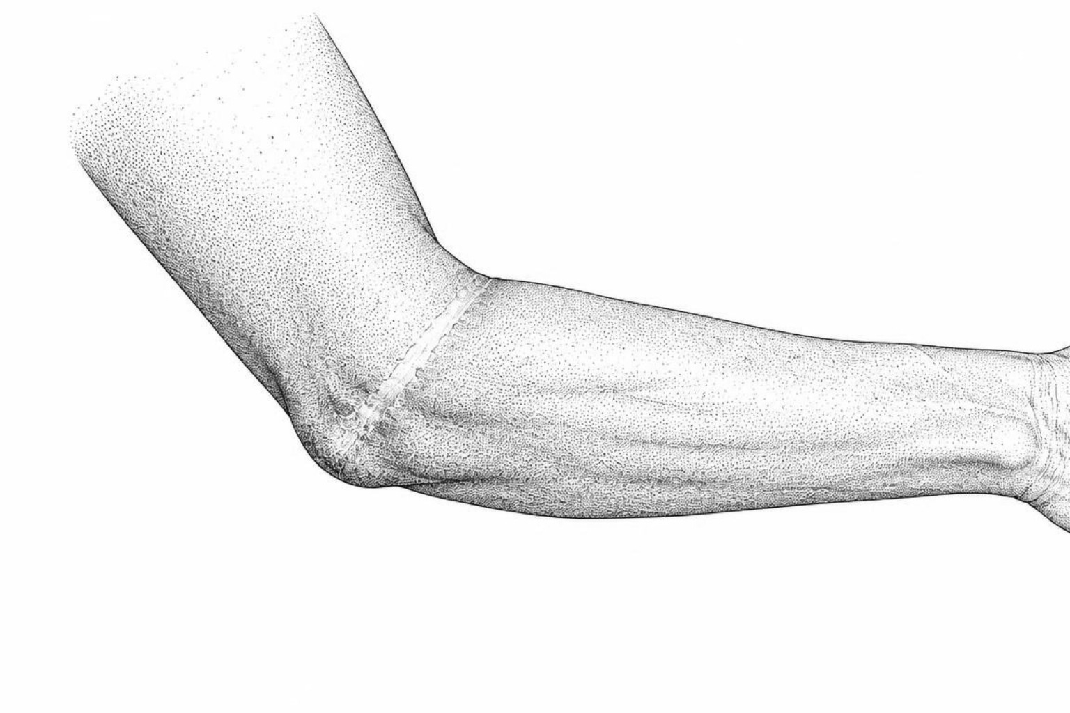
Contracture scars tighten over time and can limit joint movement. This guide explains why they persist, which early steps slow them and when splinting, laser or surgery becomes necessary.
Do contracture scars go away?
A scar that feels tight across a joint and reduces movement is unlikely to fully go away on its own. Contracture scars can soften during the first months of healing, but an established scar band often needs guided treatment to prevent lasting stiffness and loss of motion.
Deep burns, surgery, trauma, or wounds that take a long time to close can leave skin with less stretch. As the scar heals, it pulls inward and may gradually restrict the nearby skin, muscles, tendons, or joint.
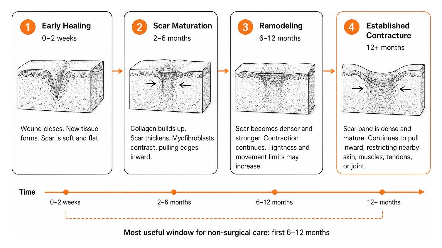
The most useful window for non-surgical care is usually within the first 6 to 12 months after the wound closes, while the scar is still changing. Daily therapy-led movement, positioning, splints, silicone, and pressure treatment may help keep the area flexible.
Book a medical assessment promptly if the scar prevents full bending or straightening of a joint, tightens week by week, causes pain with movement, or affects everyday tasks such as walking, dressing, or using a hand. The Scar Healing Editorial Team outlines why early support matters and what treatment can realistically achieve.
Why contracture scars stick around
Established contracture scars do not resolve spontaneously without structured clinical intervention. When skin sustains a full-thickness injury, the body does not replace the lost surface area with identical, elastic tissue. Instead, it synthesizes a rigid extracellular matrix composed of disorganized collagen fibers designed to achieve rapid wound closure.
Over time, this newly formed tissue contracts. While superficial scars often flatten and soften over a period of 12 to 18 months, contracture scars involve a physical deficit of cutaneous tissue. Because less surface area exists than prior to the injury, mechanical tension pulls adjacent healthy tissue taut. When this occurs across mobile anatomical structures such as the neck, wrist, elbow, or digits, the scar acts as an unyielding tether. Allowing a contracture to heal without clinical guidance permits progressive tissue shortening, converting transient movement restrictions into fixed postural deformities.
What keeps scar tissue tight
Understanding how scars form at a cellular level helps explain why a contracted scar resists natural resolution. During the proliferative and remodeling phases of wound repair, specialized myofibroblasts populate the wound bed. These cells contain contractile actin microfilaments similar to those found in smooth muscle. Myofibroblasts anchor to the extracellular matrix and physically pull wound margins inward to minimize surface area.
In standard physiologic healing, myofibroblasts undergo programmed cell death once epithelialization completes. In deep burns, extensive trauma, or infected wounds, sustained mechanical tension maintains myofibroblast activity over prolonged intervals. These cells continue depositing dense bundles of collagen, causing fibrotic bands to mature into permanent structures. Raborn and Janis describe splinting, targeted stretching, and positioning as core prevention strategies because healing tissue contracts when external positioning does not counter the pull.
The best time to treat contracture scars
Scar tissue remains biologically active and responsive to mechanical input for a limited duration. During the initial 6 to 12 months following wound closure, collagen turnover remains elevated. Applying continuous tension, controlled thermal modalities, and compressive forces during this period encourages newly deposited collagen fibers to align in parallel bundles, improving tissue pliability.
Once a scar fully matures, typically between 12 and 18 months, dense collagen cross-linking stabilizes the fibrotic band into a rigid structure. At this stage, conservative measures lose mechanical leverage. Masanovic and Téot describe early mechanical remodeling as important because mature fibrotic bands do not lengthen through manual stretching alone.
When contractures need medical care
Contracture scars represent functional movement disorders in addition to structural cutaneous changes. Managing these lesions without clinical oversight presents substantial risks to long-term joint function and structural alignment.
How contractures limit joint movement
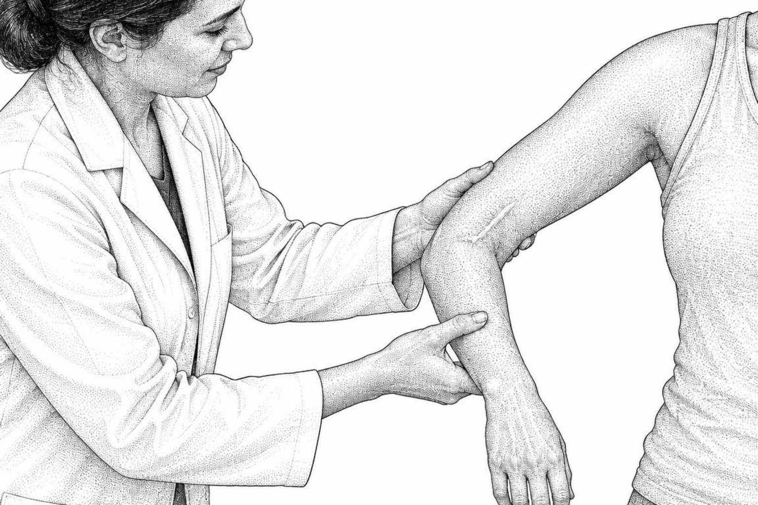
The primary clinical concern in scar contracture is progressive shortening of the skin surface. When deep tissue trauma heals across an articulation, wound margins pull tissue from adjacent anatomical zones. If an affected joint remains immobilized in flexion to minimize tension during recovery, the healing tissue consolidates that shortened posture.
Care protocols for second-degree burns demonstrate that early positioning protocols are vital because contractile forces operate continuously. Raborn and Janis report a clinical study of 145 patients with burn injuries across multiple joints in which 58.6% of operated joints showed limited range of motion at 3 to 6 weeks, dropping to 20.9% at 12 months when structured rehabilitation programs were carried out. Without clinical guidance to maintain extension through customized orthoses, skin surface deficiency progressively diminishes the functional movement arc.
Joint stiffness and tendon problems
Cutaneous contractures do not remain restricted to superficial layers. Chronic external tension from shortened skin alters the biomechanics of underlying deep structures, including muscles, tendons, joint capsules, and ligaments. When skin tightness immobilizes a joint for months, the internal joint capsule undergoes secondary shrinkage and intra-articular fibrosis.
Masanovic and Téot describe chronic extra-articular skin tension as a cause of permanent joint stiffness, muscle atrophy, and tendon adherence in hand contractures. Clinicians utilize passive motion testing to differentiate isolated cutaneous restriction from deep capsular contracture. Delayed intervention often means that releasing the cutaneous scar alone is insufficient, necessitating secondary capsulectomies or tenolyses to restore joint mobility.
Who treats contractures and how
Managing contracture pathology requires precise clinical assessment that cannot be achieved with generic stretching regimens or topical cosmetic agents. Therapeutic success depends on evaluating the dimensional vector and anatomical depth of the lesion. Linear contractures behave differently from broad, three-dimensional planar contractures that adhere to underlying deep fascia.
Medical specialists evaluate scar architecture to determine whether targeted physical therapy suffices or if surgical tissue rearrangement is indicated. Clinical classification of contracture scars enables clinicians to categorize these formations accurately and select appropriate combinations of dynamic splinting, energy-based therapies, and surgical releases.
Rehab and surgical options
Treating contracture scars involves multidisciplinary care designed to preserve motion, soften fibrotic tissue, and restore missing skin area. A general treatments overview can help readers compare clinical modalities after a clinician has measured joint motion.
Early home care and lasers
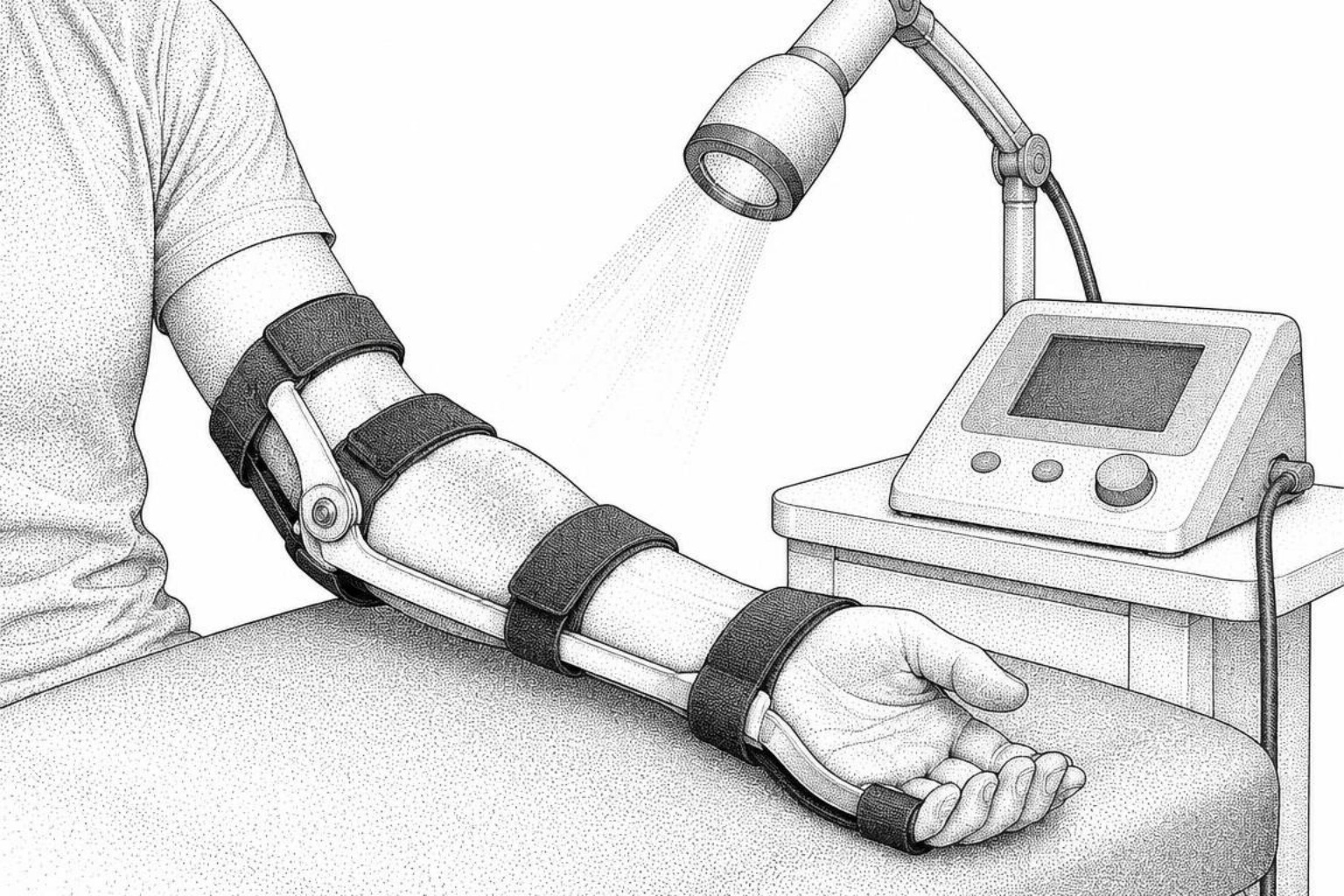
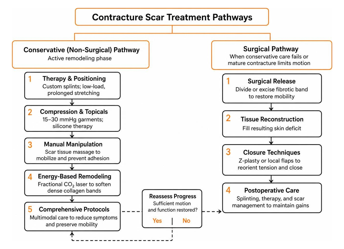
Non-surgical management forms the frontline defense during the active remodeling phase. Clinicians combine positioning, targeted stretching, compression, and topical agents to guide tissue alignment:
- Physical and Occupational Therapy: Therapists design custom dynamic and static splints to maintain joints in full extension or functional flexion. Patients perform low-load, prolonged stretches several times daily to encourage tissue lengthening.
- Compression and Topical Barriers: Custom compression garments, often in the 15 to 30 mmHg range, may reduce tissue edema and restrict excessive collagen synthesis. Using silicone treatments underneath garments provides hydration and cellular signaling that helps soften rigid scars.
- Manual Manipulation: Structured scar tissue massage may help mobilize cutaneous layers and prevent deep tissue adhesions once wounds are fully closed.
- Energy-Based Remodeling: Advanced modalities outlined in the fractional CO2 laser guide can soften dense collagen bands through microscopic thermal treatment zones. Basson and Bayat described a clinical trial of 45 patients in which four to six sessions of fractional CO2 laser yielded a 30% to 50% improvement in scar pliability, serving as a non-surgical adjunct or prehabilitation step before surgical release.
- Comprehensive Burn Protocols: Protocols outlined in the guide for non-surgical burn scar care emphasize using continuous multimodal therapies to manage symptoms like neuropathic itching while preserving tissue mobility.
Surgical release and reconstruction
When a mature contracture locks a joint or conservative therapy fails to restore range of motion, surgical intervention is required. The surgical objective is to divide or excise the fibrotic band and fill the resulting skin deficit:
- Z-Plasty and Local Flaps: For linear contractures, surgeons use Z-plasty transposition flaps. Masanovic and Téot describe a standard 60-degree Z-plasty as a technique that reorients the tension line by 90 degrees and yields an approximate 75% gain in functional tissue length along the contracture line.
- Skin Grafts: Full-thickness skin grafts (FTSG) are placed over released tissue beds where local skin is unavailable. Full-thickness grafts resist secondary contraction significantly better than thin split-thickness skin grafts.
- Fasciocutaneous and Free Flaps: For deep or planar contractures, surgeons transfer vascularized tissue flaps. Raborn and Janis reviewed 327 studies and noted that flap reconstruction demonstrates lower rates of contracture recurrence compared to skin grafts, particularly over high-mobility joints like the neck and elbow.
- Surgical Revision Planning: The scar revision treatment guide highlights how combining surgical releases with post-operative splinting prevents the reconstructed site from reforming contracture bands during new healing phases.
Common questions about contracture scars
Can a contracture scar be fully removed?
A contracture scar cannot be erased entirely. Adult skin heals by forming scar tissue, so surgical interventions replace a tight, restrictive scar with a longer, more flexible, and strategically positioned surgical line. The primary goal of treatment is the full restoration of joint mobility, pain relief, and tissue softening. In a scar management review, Khansa, Harrison, and Janis describe scar recovery as a process that depends on matching the procedure to the scar and following it with diligent post-procedure therapy to improve functional range of motion and joint mobility.
When to get care after a burn or injury
Rehabilitation and medical assessment should begin during the active healing phase, before full wound closure occurs. Gentle range-of-motion exercises and proper resting positioning prevent the initial formation of restrictive webs. Once re-epithelialization is complete, formal therapy involving splinting, compression, and silicone applications should be initiated immediately to take advantage of the early 6 to 12 month remodeling window.
Does insurance cover contracture release?
Health insurance plans generally cover the evaluation, physical therapy, and surgical release of contracture scars when medical necessity is documented. Coverage criteria depend on demonstrable functional limitations, such as restricted joint angles, chronic pain, or inability to perform activities of daily living. Detailed range-of-motion measurements, clinical photographs, and occupational therapy assessments provide the necessary documentation for insurance approval.
What to expect from a contracture scar
Contracture scars represent complex structural changes where lost skin surface area exerts constant mechanical pull on joints and deeper soft tissues. These scars do not resolve on their own, and leaving them unmanaged risks permanent joint capsule stiffness and chronic movement restriction. Achieving long-term functional recovery requires early, structured intervention combining physical therapy, dynamic splinting, advanced laser remodeling, and surgical reconstruction when necessary.
For an individualized evaluation of your scar tissue and functional mobility, complete a comprehensive scar assessment to determine the appropriate clinical next steps.
Works Cited
- Masanovic MG, Téot L. "Scar Contractures." Springer, 2020.
- Raborn LN, Janis JE. "Prevention and Treatment of Burn Scar Contracture: A Practical Review." Plastic and Reconstructive Surgery Global Open, 2024.
- Basson R, Bayat A. "Skin scarring: Latest update on objective assessment and optimal management." Frontiers in Medicine, 2022.
- Khansa I, Harrison B, Janis JE. "Evidence-Based Scar Management: How to Improve Results with Technique and Technology." Plastic and Reconstructive Surgery, 2016.
This content is for informational purposes only and does not constitute medical advice. Consult a qualified healthcare professional for diagnosis and treatment.