A Practical Guide to Why Two People Scar Differently: Formation Factors
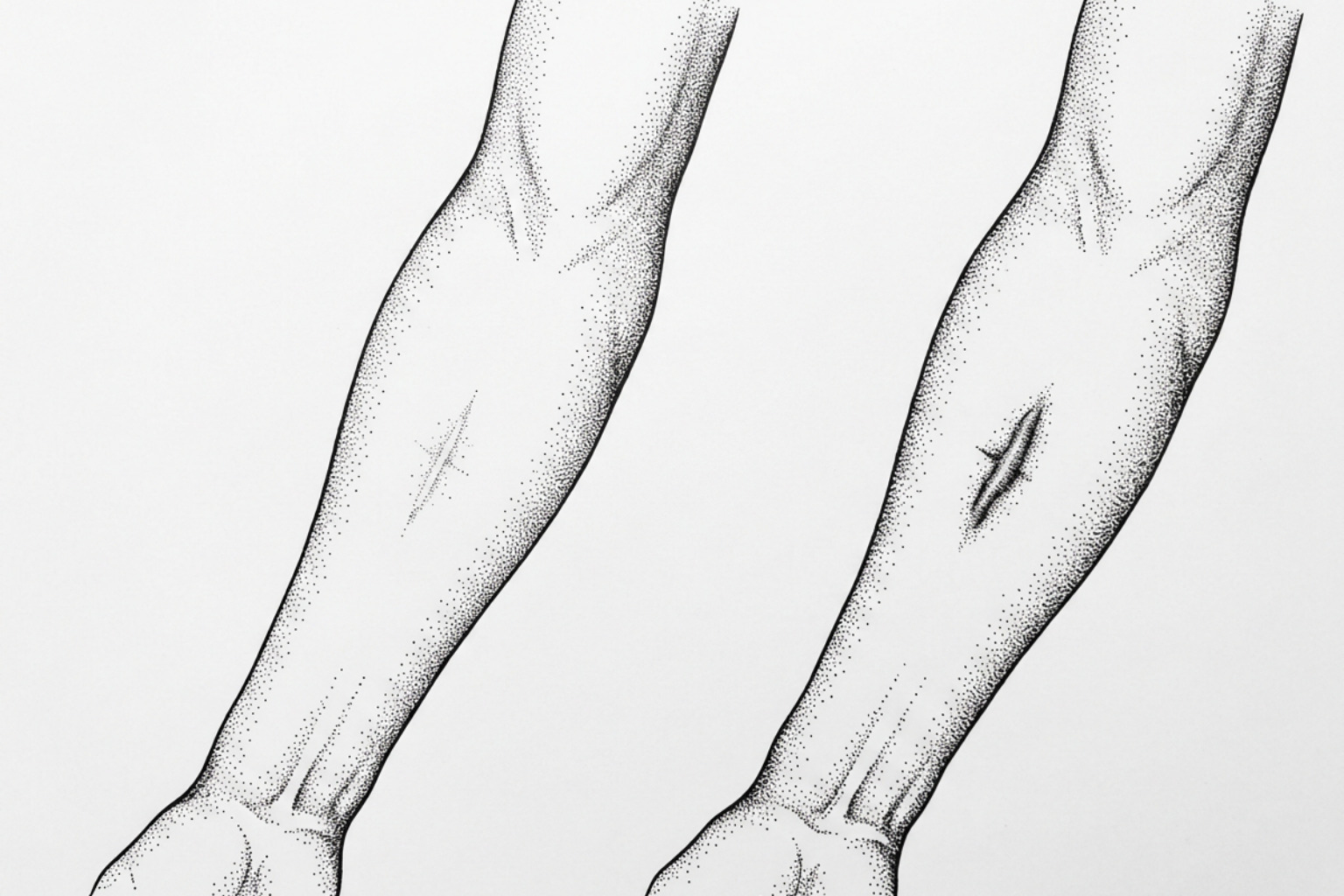
Two people can get the same cut and end up with very different scars. Genetics, fibroblast behaviour, skin tone and the tension across the wound all play a part — and some of those factors you can influence.
Why scars differ from person to person
Why two people scar differently comes down to a mix of biology, the wound itself, and the forces acting on healing skin. Even when two people have the same surgery or injury, their bodies do not repair tissue in exactly the same way.
Research indicates that scar outcomes can differ because of:
- Genetics and immune response: Genes and inflammatory signals can affect how strongly fibroblasts - the cells that build collagen - respond after injury.
- Skin tone and pigment activity: Melanin-rich skin may be more prone to lasting dark marks after inflammation and, in some people, raised scars such as keloids.
- Wound depth, infection, and healing time: Deeper wounds and wounds that stay open longer have a higher risk of abnormal or raised scarring.
- Body location and skin tension: Areas that stretch or move often, including the chest, shoulders, and joints, can place more mechanical stress on a healing wound.
- Age, health, and lifestyle factors: Conditions that affect circulation or inflammation, as well as smoking and poor nutrition, may alter tissue repair.
A scar is the body’s fast repair system. Fibroblasts lay down collagen, a strong structural protein, to close damaged skin. This protects the body, but the repaired area may not rebuild normal features such as hair follicles, sweat glands, or the original pattern of collagen fibers.
Evidence also suggests that scars are not only about appearance. Itch, pain, tightness, color change, and self-confidence can vary widely between people. Standard clinical scar scales do not always capture those differences equally across skin types, which is one reason personal experience matters alongside an in-person assessment.
The Scar Healing Editorial Team examines the science behind these differences, starting with the biological signals that steer a wound toward a faint line, a dark mark, a depressed scar, or a raised one.
The biology and genetics behind it
When skin experiences an injury, a precise biological sequence begins: hemostasis stops bleeding, acute inflammation clears cellular debris, proliferation builds new dermal tissue, and maturation slowly remodels the extracellular matrix (ECM). To understand How Scars Form, it helps to observe that this sequence is governed by deep genetic programming. Two individuals receiving identical surgical incisions may activate entirely different genomic pathways during these phases.
Genes linked to heavy scarring
Genome-wide association studies (GWAS) show that underlying genetic variations heavily influence whether a wound forms a fine, flat line or a thick, fibrotic scar. Research on genomic factors in scarring has identified specific single-nucleotide polymorphisms (SNPs)—tiny variations in DNA sequences—that predispose certain individuals to aggressive fibrotic responses.
For example, specific SNP loci such as rs873549, rs940187, rs1511412, and rs8032158 near the FOXL2 and NEDD4 gene regions strongly correlate with keloid susceptibility. Furthermore, loss-of-function variants in the MC1R gene (such as the R163Q SNP) show significant prevalence in specific populations; allele frequencies reach approximately 70% in East and Southeast Asian cohorts and up to 100% in Native American populations. These variants alter cutaneous signaling pathways, predisposing individuals to heightened inflammatory responses and hypertrophic scar development.
Fibroblasts and the signals they follow
Fibroblasts are not a single, uniform population of cells. Modern single-cell sequencing reveals marked heterogeneity among dermal fibroblasts, primarily divided into papillary fibroblasts (located in the upper dermis) and reticular fibroblasts (located in the deep dermis).
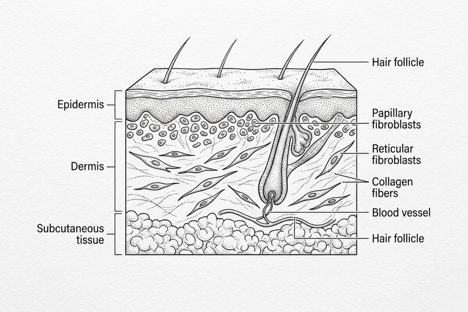
When an injury penetrates into the deep reticular dermis, reticular fibroblasts predominate. These cells exhibit lower baseline collagenase activity but secrete significantly higher levels of pro-fibrotic signaling cytokines, particularly transforming growth factor-beta 1 (TGF-β1). Under persistent inflammatory signals, reticular fibroblasts convert into myofibroblasts—specialized cells expressing alpha-smooth muscle actin (α-SMA) that generate high contractile forces and overproduce dense Type I collagen.
In addition, immune system dynamics play a pivotal role. A systemic shift toward a Th2 immune cell response (characterized by elevated interleukin-4, interleukin-10, and TGF-β) sustains myofibroblasts and prevents normal cell apoptosis, causing continuous tissue deposition rather than quiet matrix remodeling.
How skin tone affects scarring
The biological interaction between skin pigmentation, inflammation, and scar maturation explains why physical symptoms and visual outcomes vary across diverse ethnic backgrounds. Melanin synthesis and fibroblast reactivity are closely linked through inflammatory cytokine pathways.
Fitzpatrick skin types and what changes
The Fitzpatrick skin phototype (FST) scale classifies skin according to pigment levels and UV radiation response. Research on scar assessment and skin type demonstrates that physical scar symptoms—specifically pruritus (itching) and hyperpigmentation—correlate directly with skin phototype.
In a comparative trial evaluating post-injury scars at 4 months, patients with Fitzpatrick skin type V (darker skin) reported significantly higher self-reported itch scores (7.3 ± 0.6) on the Patient and Observer Scar Assessment Scale (POSAS) compared to patients with Fitzpatrick skin type II (5.3 ± 0.7, p < 0.05). Spectrophotometric evaluations revealed that melanin levels were significantly higher in FST-V scars (896.4 ± 9.5) compared to FST-II scars (727.2 ± 9.8, p < 0.0001). Conversely, lighter skin types (FST-II) exhibited higher erythema (redness) values (463.7 ± 6.4) than darker skin types (398.7 ± 11.4, p < 0.0001). Understanding Why Scars Itch requires recognizing that elevated melanin activity correlates with heightened neurogenic inflammatory signaling in the dermis.
| Fitzpatrick Skin Type | Predominant Optical Feature | Mean Melanin Index | Mean POSAS Itch Score | Primary Symptom Burden |
|---|---|---|---|---|
| FST-II (Lighter Skin) | High Erythema (463.7 ± 6.4) | 727.2 ± 9.8 | 5.3 ± 0.7 | Persistent Redness, Telangiectasia |
| FST-V (Darker Skin) | High Melanin (896.4 ± 9.5) | 896.4 ± 9.5 | 7.3 ± 0.6 | Pruritus, Hyperpigmentation |
Who gets keloids most often
Clinical observations show striking disparities in severe fibrotic scar development. African American patients exhibit a 15- to 20-fold greater statistical risk of keloid formation compared to individuals with lower baseline pigmentation. A comprehensive review of Keloid Causes Complete Guide highlights that keloid incidence reaches up to 16% in populations of African and Hispanic ancestry.
These physiological differences directly influence patient quality of life. In a multi-center study using the validated SCAR-Q outcome tool, African American surgical patients reported significantly lower overall satisfaction scores (203 ± 116) compared to White patients (232 ± 79, p < 0.001). Longitudinal tracking further revealed a divergence in recovery trajectories: while White patients showed statistically significant improvements in scar symptoms and appearance over time (r = 0.18, p < 0.001), African American patients demonstrated no statistically significant symptom improvement over time (p = 0.11 for symptoms, p = 0.37 for appearance). This persistence of physical symptoms contributes to documented psychosocial stress, body image self-consciousness, and daily discomfort.
Why the place on your body matters
The physical location of a wound introduces distinct developmental and mechanical variables. Wounds located on the face typically heal with finer, less noticeable scars than identical wounds located on the trunk or extremities.
Why facial wounds scar less
A key reason why why two people scar differently across body sites stems from embryonic cell lineage. Facial dermal tissue originates from embryonic neural crest cells, whereas body skin arises from mesodermal lineages. A landmark Stanford study on facial scar resistance revealed that facial fibroblasts maintain a progenitor-like state through active ROBO2 protein signaling.
This ROBO2 pathway keeps DNA transcriptionally less accessible for pro-fibrotic collagen genes. In experimental models, activating ROBO2 signaling in just 10% to 15% of fibroblasts surrounding a body wound was sufficient to transform the repair response, causing back wounds to heal with the reduced scarring characteristic of facial skin. Furthermore, small-molecule inhibition of EP300 (an epigenetic regulator) suppressed pro-fibrotic gene activation, steering adult skin away from heavy fibrosis toward low-scar tissue repair.
How skin tension shapes a scar
Mechanical forces play a powerful role in scar biology through mechanotransduction—the process by which living cells sense and translate physical tension into intracellular biochemical signals.
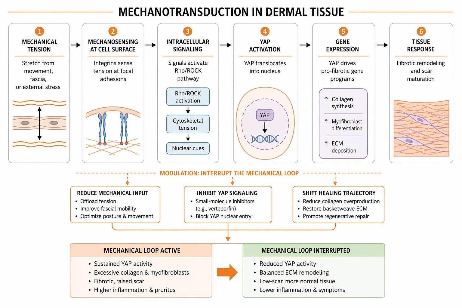
In regions under continuous dynamic stress (such as the chest, shoulders, or joint surfaces), skin tension activates the YAP (Yes-associated protein) mechanotransduction pathway. Physical tension causes YAP to translocate into the fibroblast nucleus, driving continuous collagen synthesis and myofibroblast differentiation.
Study on molecular trajectories in scarring demonstrates that blocking YAP signaling (for example, with small-molecule agents like verteporfin) interrupts this mechanical loop, shifting wound repair from a fibrotic path to a regenerative trajectory that restores normal basketweave ECM architecture and functional skin appendages.
The fascial network and central nervous system further modulate this environment; deep tissue adhesions disrupt tissue layer movement, while central nervous system stress responses elevate systemic cortisol, prolonging inflammatory signaling and altering wound tension.
S.C.A.R. endotypes, and what scales miss
Historically, clinicians classified scars purely by their surface appearance (phenotype). However, contemporary dermatology recognizes that visible features are driven by underlying biological mechanisms known as endotypes.
How the S.C.A.R. model groups scars
To standardize clinical evaluation, Research on dermal fibrosis endotypes established the S.C.A.R. endotype framework. This model categorizes human dermal fibrosis into four primary biological pathways, enabling precise diagnostic alignment:
- Stretched (Flat): Characterized by dermal collagen thinning without excessive elevation; frequently driven by low-grade mechanical strain across Langer's lines.
- Contracted: Characterized by tight, restrictive tissue bands causing structural shortening; predominantly driven by persistent myofibroblast α-SMA contraction following extensive deep dermal injury.
- Atrophic (Depressed): Characterized by localized dermal tissue loss and sunken contours; caused by matrix degradation, insufficient collagen synthesis, or underlying adipose loss.
- Raised (Hypertrophic and Keloid): Characterized by excessive, continuous extracellular matrix accumulation; driven by persistent reticular fibroblast activation, high TGF-β signaling, and attenuated collagenase turnover.
Standardized clinical Scar Assessment relies on identifying these specific endotypes rather than treating all raised or discolored marks identically.
Where standard scar scales fall short
Traditional scar assessment scales—such as the Vancouver Scar Scale (VSS) and the Patient and Observer Scar Assessment Scale (POSAS)—rely heavily on subjective clinician ratings of height, vascularity, and pliability. Recent clinical evaluations reveal that these tools exhibit notable limitations across diverse Fitzpatrick skin types.
While observers using VSS or POSAS-O record similar numerical scores for scars on light (FST-II) and dark (FST-V) skin (POSAS-O: 6.2 ± 0.4 vs. 6.1 ± 0.4), these subjective scales fail to capture underlying physiological variations such as precise melanin density or neurogenic itch severity. To overcome these limitations, modern dermatological research incorporates objective, non-invasive measuring devices, including spectrophotometers for melanin/erythema quantification, tissue durometers for elasticity, and high-frequency ultrasound to assess deep dermal collagen thickness.
How to lower your own scarring risk
Because individual scar trajectories vary widely, evidence-based management emphasizes early intervention, precise mechanical control, and protocols matched to specific risk profiles.
Early care and simple barrier treatments
The window immediately following re-epithelialization is critical for long-term scar maturation. Wounds that take longer than 21 days to achieve complete epithelial closure carry an elevated hypertrophic scarring risk of up to 78% in adults (compared to 33% for wounds closing within 14 to 21 days).
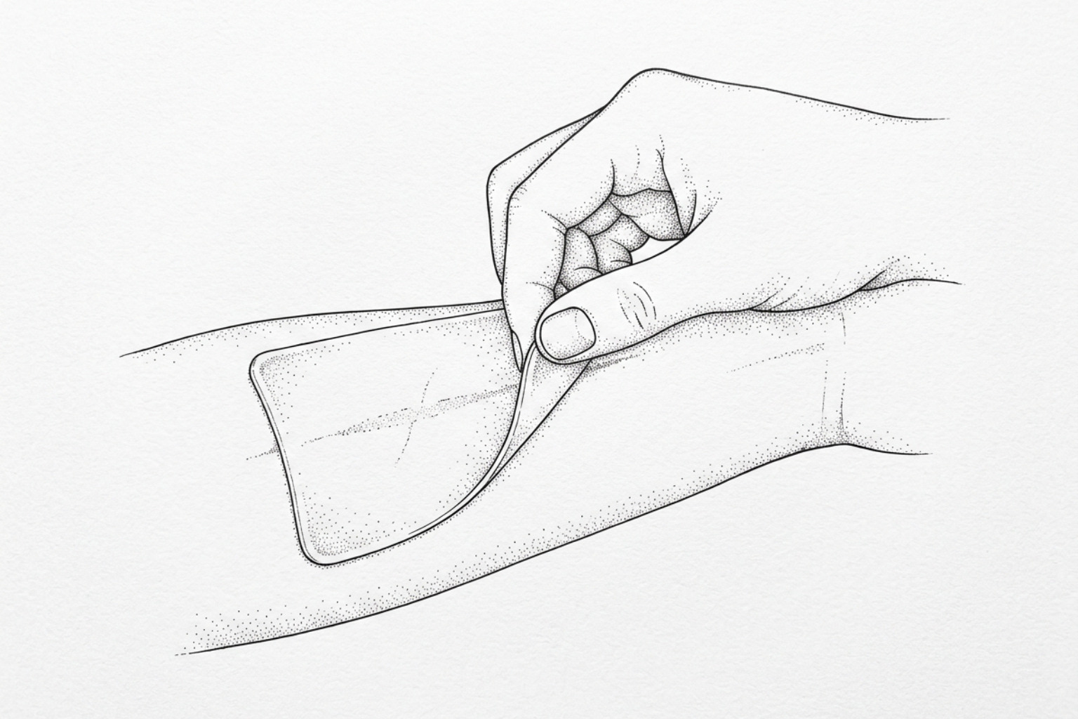
Topical silicone barrier therapies—available as sheets or self-drying gels—represent established first-line non-invasive management according to international clinical consensus guidelines (Level I clinical evidence). Systematic reviews of randomized trials indicate moderate efficacy in reducing scar thickness and pruritus; however, patient compliance can be hindered by localized skin maceration, contact dermatitis, or difficulty maintaining occlusion on high-mobility joints. Silicone restores stratum corneum hydration, reducing transepidermal water loss (TEWL). This hydration signal sends direct feedback to superficial keratinocytes, which in turn release stratifin (14-3-3σ), an anti-fibrogenic protein that downregulates pro-fibrotic collagen synthesis in underlying dermal fibroblasts. Adhering to structured Dermatology Scar Management Protocols during early matrix assembly helps normalize local collagen alignment.
When to consider advanced treatment
For high-risk or recalcitrant scars, second- and third-line interventions target distinct cellular mechanisms, though each carries specific levels of evidence, variable response rates, and potential adverse effects:
- Intralesional Corticosteroids: Injections of triamcinolone acetonide suppress fibroblast proliferation, inhibit TGF-β1 expression, and promote matrix metalloproteinase production. Meta-analyses of clinical trials (Level II evidence) demonstrate objective response rates between 50% and 100%. However, limitations include injection discomfort, localized skin atrophy, telangiectasia, post-inflammatory hypopigmentation (particularly in melanin-rich skin), and reported symptom recurrence rates up to 50% after treatment cessation.
- Pulsed Dye Laser (595 nm): Selectively targets microvascular hemoglobin, inducing selective photothermolysis of persistent capillary beds to reduce erythema and tissue perfusion. A randomized controlled trial of 30 participants evaluated 595 nm pulsed dye laser therapy and demonstrated moderate reductions in scar redness and height (Level II evidence). Limitations include limited effect on dense hyperplastic collagen, the need for multiple treatment sessions, significant financial cost, and risks of transient purpura or secondary hyperpigmentation.
- Fractional Ablative Laser Therapy: Creates microscopic thermal zones in dense scar tissue, triggering a localized wound healing response that degrades disorganized collagen bundles. A prospective study of 42 surgical and burn patients demonstrated measurable improvements in scar pliability and texture (Level II/III evidence). Key limitations include prolonged healing downtime, procedure discomfort, risk of thermal injury, and potential post-inflammatory hyperpigmentation. Understanding How to Improve Collagen Remodeling in Scars requires combining appropriate thermal or mechanical stimulation with strict post-procedure barrier care.
Emerging Precision Scar Medicine (PSM) utilizes biobanking, genomic sequencing, and proteomic biomarker profiling to predict scar risk prior to elective procedures, allowing clinicians to tailor antifibrotic therapies directly to an individual's biological profile. While early observational cohort data support this personalized approach, large-scale multi-center randomized controlled trials remain necessary to confirm long-term clinical utility and cost-effectiveness.
Frequently asked questions
Why do wounds on the face heal with significantly less scarring than body wounds?
Facial skin cells derive from embryonic neural crest cells, whereas body skin cells originate from mesoderm. Research indicates that facial fibroblasts maintain higher active ROBO2 signaling, keeping their chromatin in a progenitor-like state that downregulates pro-fibrotic collagen genes and promotes a regenerative repair response.
Why are individuals with darker skin tones more susceptible to keloid scars?
Individuals with darker skin tones (Fitzpatrick skin phototypes IV–VI) possess heightened melanocyte activity and specific genetic susceptibilities, including key SNP loci near FOXL2 and NEDD4. These factors drive stronger reticular fibroblast activation, prolonged inflammatory signaling, and a 15- to 20-fold increased statistical risk of keloid formation compared to lighter skin types.
Can personalized medicine predict how an individual will scar before surgery?
Yes, advancing Precision Scar Medicine uses next-generation whole-genome sequencing and systemic biomarker screening to identify high-risk genetic variants (such as MC1R or FOXL2 mutations) prior to elective surgery. This allows clinicians to implement targeted preventative protocols immediately upon wound closure.
What this means for you
Understanding why two people scar differently highlights the complex interplay between genomic profiling, cellular signaling, skin phototype, mechanical forces, and anatomical location. Fibrotic tissue repair is not a uniform process, but rather a spectrum of biological responses ranging from minimal tissue alteration to extensive keloids and contractures.
As research continues to unravel the molecular mechanisms of dermal repair—from YAP mechanotransduction to neural crest fibroblast lineages—scar management is moving away from generalized approaches toward individualized care. By incorporating objective diagnostic measurement, recognizing unique skin phototype needs, and utilizing early targeted interventions, clinicians can optimize physical and visual scar outcomes for every individual. For an in-depth review of tailored strategies for melanin-rich skin, consult our Comprehensive Guide to Scar Management in Darker Skin Types.
This content is for informational purposes only and does not constitute medical advice. Consult a qualified healthcare professional for diagnosis and treatment.
Works Cited
- Borsese A, et al. "Are There Differences in Scar Assessments as a Function of Fitzpatrick Skin Type? (abstract 129)." Journal of Burn Care & Research, 2024;45(Suppl 1):i98-i99.
- Conger K. "The face scars less than the body - a Stanford Medicine study unravels why." Stanford Medicine News, 2026.
- Mascharak S, et al. "Multi-omic analysis reveals divergent molecular events in scarring and regenerative wound healing." Cell Stem Cell, 2022;29(2):315-327.e6.
- Ud-Din S, Bayat A. "Classification of Distinct Endotypes in Human Skin Scarring: S.C.A.R. - A Novel Perspective on Dermal Fibrosis." Advances in Wound Care, 2022;11(3):109-120.
- Amini-Nik S. "Time Heals all Wounds - but Scars Remain. Can Personalized Medicine Help?." Frontiers in Genetics, 2018;9:211.
- Xue M, Jackson CJ. "Extracellular Matrix Reorganization During Wound Healing and Its Impact on Abnormal Scarring." Advances in Wound Care, 2015;4(3):119-136.