Stretch Marks on Back: Growth Spurts and Skin Care
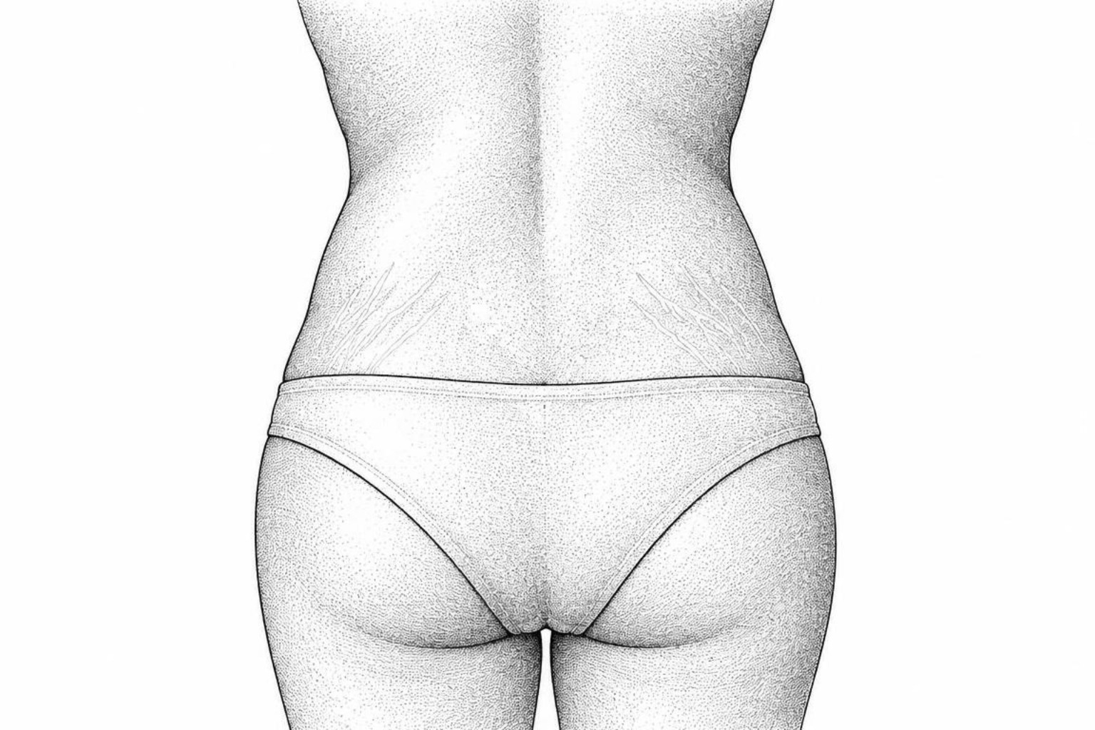
Stretch marks on the back often appear during a growth spurt, weight gain or fast muscle growth. Learn what causes them, how they fade from red to pale, and what moisturizers and in-office treatments can do.
Why stretch marks appear on the back
Stretch marks on back skin often form during a growth spurt, rapid weight gain, or fast muscle growth, leaving a small scar where the dermis could not keep pace with the stretch. Early management emphasizes maintaining epidermal hydration to ease localized pruritus and avoiding abrupt fluctuations in body mass.
The lesions develop when underlying tissue expands faster than supporting dermal fibers can adapt. Initial marks present as erythematous, violaceous, or hyperpigmented bands, and many gradually fade over 6 to 12 months, although dermal scarring rarely resolves entirely. Consistent hydration can relieve surface dryness, but topical emollients cannot eliminate established structural scarring.
Patients should seek medical evaluation if wide, dark-purple striae appear suddenly alongside easy bruising or proximal muscle weakness. Therapeutic discussions frequently explore modalities such as lasering stretch marks, mechanical strain mechanisms observed in pregnancy and stretch marks, and the clinical utility of a targeted stretch marks treatment cream. The Scar Healing Editorial Team focuses on practical skin care and realistic expectations.
What causes back stretch marks
The skin possesses natural elasticity, yet rapid mechanical tension can overwhelm its structural integrity. The dermis, which serves as the middle structural layer of the skin, consists of collagen bundles that supply firmness and elastin networks that provide rebound resilience. When the body undergoes rapid physical expansion, dermal fibroblasts cannot produce extracellular matrix components fast enough to accommodate the mechanical strain.
This mechanical force leads to microscopic ruptures within the dermal connective tissue. As the elastin mesh breaks, the superficial blood vessels become visible through the thinned dermal matrix, producing the initial inflammatory color. Over time, the body deposits dense, disorganized scar collagen to repair the micro-tears, creating the classic linear texture known medically as striae distensae.
Growth spurts in teens
During peak adolescent development, skeletal elongation occurs rapidly. When the spine lengthens faster than the overlying dermis can expand, perpendicular mechanical stress pulls the skin taut. This vertical spinal growth creates distinctive horizontal linear marks across the lumbar or thoracic spine.
In a study of 12 teenage boys with horizontal stretch marks on the lower back, two thirds reported a significant growth spurt before the marks appeared, and 11 of the 12 had a close relative with stretch marks (Boozalis et al., 2018). These marks often resemble the rungs of a ladder, spanning evenly across the lower spine. The same study found no link to chronic illness, infection or steroid use, so this pattern reflects normal physiological development and does not indicate external skin trauma.
Weightlifting and muscle growth
Intensive resistance training produces rapid increases in skeletal muscle volume. Bodybuilders and strength athletes often experience localized skin stretching over the latissimus dorsi, trapezius, and posterior deltoids. When high-volume training cycles cause muscle bellies to expand faster than dermal remodeling can occur, tensile stress concentrates in the upper back.
Adopting a gradual progression in training volume and maintaining steady caloric surpluses allows the dermal architecture to adapt to mechanical strain, reducing the severity of acute dermal tearing.
Hormones and medical conditions
Hormones directly regulate structural skin integrity. Elevated glucocorticoids, whether from internal physiological stress, external medications, or systemic endocrine disorders, inhibit dermal fibroblasts from synthesizing new collagen and elastin. Prolonged systemic or high-potency topical corticosteroid use weakens the dermal matrix, making the skin susceptible to wide striae under minimal physical tension.
In endocrine conditions such as Cushing syndrome, sustained cortisol excess thins the dermal layer and produces prominent, violaceous striae that measure wider than standard growth marks. Connective tissue disorders like Marfan syndrome and Ehlers-Danlos syndrome alter collagen synthesis genetically, allowing striae to form during minor physical shifts. Distinct morphological signs, such as broad purple bands or systemic skin laxity, warrant medical evaluation.
What back stretch marks look like
Evaluating the orientation, width, and color of back marks helps identify their origin and timeline.
| Characteristic | Horizontal Growth Lines | Vertical Tension Lines |
|---|---|---|
| Primary Location | Lower back (lumbar spine), mid-thoracic region | Upper back, latissimus dorsi, rear shoulder area |
| Primary Trigger | Rapid vertical height growth during puberty | Rapid muscle hypertrophy or broad torso expansion |
| Visual Pattern | Parallel horizontal lines resembling ladder rungs | Perpendicular, vertical or diagonal streaks |
| Initial Appearance | Pink, red, or light purple thin bands | Erythematous, slightly raised ridges |
| Mature Stage | Silvery-white, flat or slightly depressed lines | Depressed, finely wrinkled, hypo-pigmented tracks |
Horizontal vs. vertical marks
Horizontal marks along the spine develop in response to vertical skeletal stretching. These marks often appear symmetrically across the lumbar curve. In some cases, chronic sedentary posture causes skin folds that compress the dermis, creating temporary postural creases. Unlike structural stretch marks, functional postural creases lack dermal scarring and resolve with core conditioning and posture changes.
Vertical or diagonal lines across the upper back occur when lateral expansion stretches the skin horizontally. This pattern is common in strength athletes whose back width increases from latissimus dorsi development.
How red marks turn white
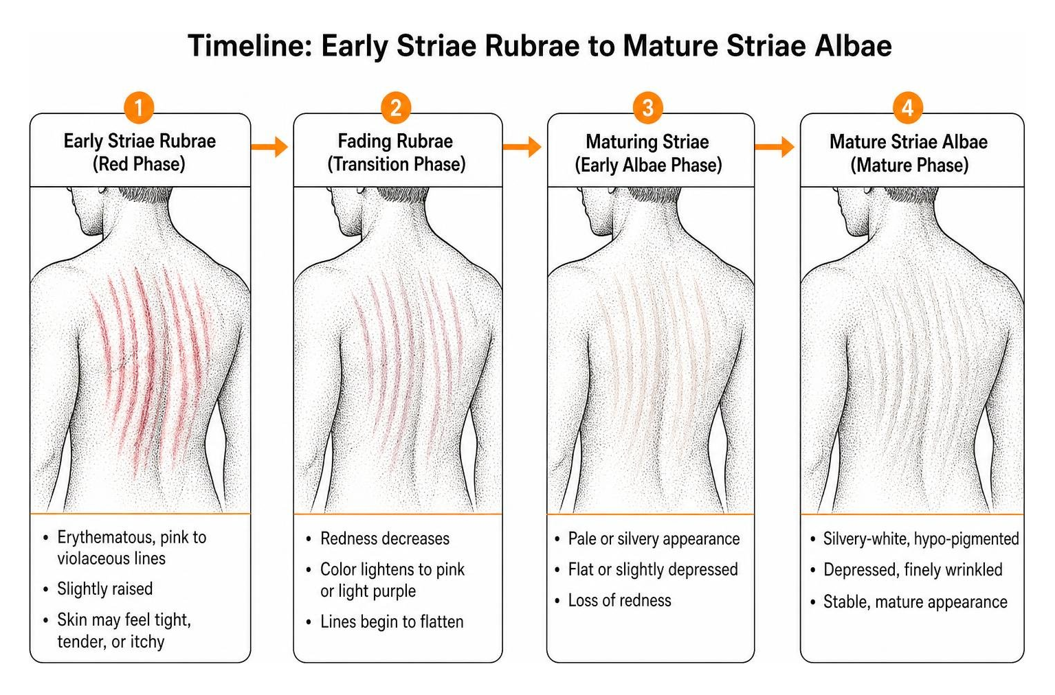
Stretch marks progress through two clinical stages:
- Striae Rubrae (Vascular Phase): Early marks appear erythematous, ranging from pink to violaceous or dark brown depending on baseline skin melanin. The overlying skin may feel tight, mildly tender, or itchy due to active mast cell degranulation and localized inflammation.
- Striae Albae (Atrophic Phase): Over 6 to 12 months, microvascular inflammation subsides and the vessels constrict. The mark flattens, loses pigmentation, and develops a slightly depressed, finely wrinkled surface.
A complete overview of stretch mark types details these histological transitions.
Creams, food and prevention
Consistent skin care supports epidermal hydration and maintains baseline skin suppleness during growth and weight shifts.
Keeping skin moisturized
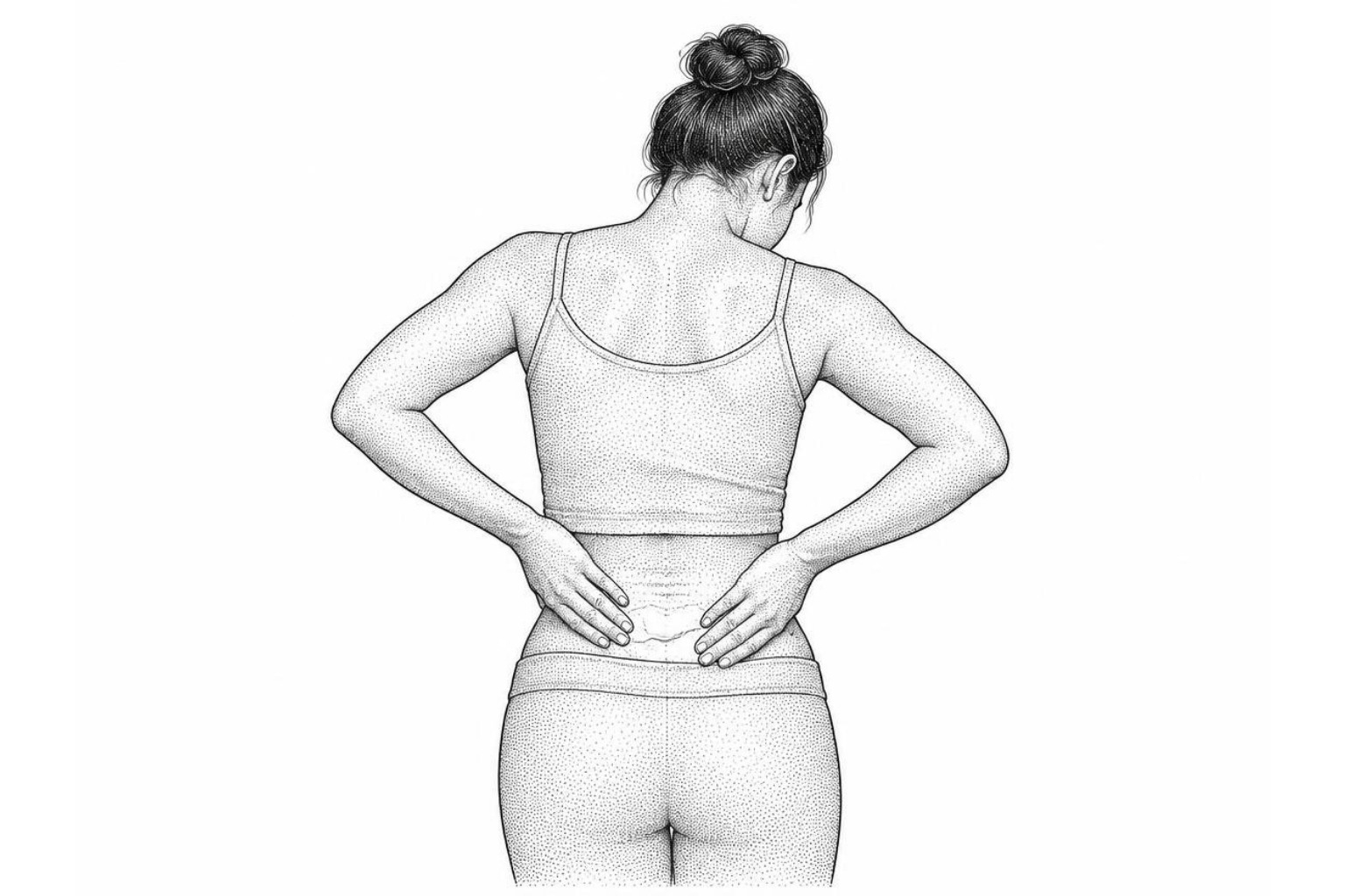
Maintaining adequate epidermal hydration minimizes transepidermal water loss and relieves tension-related itching. Humectants like hyaluronic acid and glycerin draw ambient moisture into the stratum corneum, while emollients support surface barrier integrity. Topical formulations containing centella asiatica extract support general skin conditioning.
Applying specialized stretch mark creams immediately after bathing seals in hydration when the skin barrier is most receptive. While rich botanical skin oils soften rough, tight areas, they cannot alter dermal scar architecture. In a randomized trial of 300 pregnant women, cocoa butter cream did not prevent stretch marks (Buchanan et al., 2010), and a review of prevention studies reached the same conclusion for olive oil (Korgavkar & Wang, 2015).
Nutrition and skin health
Dermal fibroblasts require specific micronutrients to synthesize structural proteins:
- Protein Intake: Supplies essential amino acids (proline, glycine, lysine) for collagen assembly.
- Vitamin C: Acts as an essential cofactor in collagen cross-linking and supports dermal resilience.
- Zinc and Vitamin D: Support cellular turnover and tissue repair mechanisms.
- Consistent Hydration: Maintains extracellular matrix volume and tissue turgor.
- Gradual Caloric Adjustments: Prevents sudden mechanical strain on the dermis from rapid weight swings.
Treatment options
For individuals seeking noticeable reduction in scar visibility, clinical dermatological modalities target collagen remodeling in the deeper dermal layers. The evidence-based stretch mark treatment guide outlines comprehensive comparative approaches.
In-office procedures
- Topical Retinoids: Prescription tretinoin promotes epidermal turnover and stimulates dermal collagen synthesis. In a randomized study of 32 women with early red stretch marks, 0.05% tretinoin cream significantly improved their appearance over 16 weeks (Hexsel et al., 2014). Retinoids can cause localized irritation and are contraindicated during pregnancy and breastfeeding.
- Microdermabrasion: Mechanical exfoliation removes superficial dead cells, smoothing the outer skin texture. In the same study, weekly superficial dermabrasion, a related and more localized abrasion technique, improved early stretch marks as much as 0.05% tretinoin cream, with fewer side effects.
- Microneedling: Fine mechanical punctures create controlled micro-injuries in the dermis, stimulating wound healing and neocollagenesis. An uncontrolled pilot study found that 7 of 16 patients (43.8%) receiving three microneedling treatments achieved marked-to-excellent visual improvement (Park et al., 2012).
- Energy-Based and Laser Therapy: Pulsed-dye lasers target the vascular components of early striae rubrae, while fractional ablative and non-ablative lasers stimulate deep dermal remodeling for older striae albae. For detailed mechanisms, review targeted laser therapy.
- Superficial Chemical Peels: Glycolic or trichloroacetic acid solutions exfoliate the epidermis and promote tissue regeneration when combined with other in-clinic treatments.
Common questions
Will back stretch marks go away?
Stretch marks represent permanent structural alterations within the dermal collagen network, so they rarely disappear entirely. However, they naturally fade over 6 to 12 months, shifting from bright red or purple to subtle silvery-white lines that blend into surrounding skin.
Can lifting cause upper back marks?
Rapid muscle hypertrophy from progressive resistance training places acute lateral tension on the overlying skin. When upper back musculature expands faster than the dermis can synthesize supporting tissue, vertical or diagonal striae can develop along the latissimus dorsi and posterior shoulders.
When to see a doctor
Back stretch marks warrant medical evaluation if they appear suddenly without clear growth or weight shifts, exceed one centimeter in width, or exhibit a deep purple hue. Prompt evaluation is also recommended if marks accompany unusual bruising, muscle weakness, systemic fatigue, or abnormal blood pressure fluctuations.
The bottom line
Back stretch marks are common, harmless responses to growth spurts, muscle building, and weight changes. Adopting realistic expectations, maintaining steady skin hydration, and managing weight gradually help keep the skin resilient. To evaluate individual skin concerns, individuals can complete a personalized scar assessment for tailored insights.
Works Cited
- Anthony K. “Stretch Marks on Back: Causes, Symptoms, and Treatment.” Healthline, 2020.
- Kandola A. “Can you get rid of stretch marks on the back? Causes and treatment.” Medical News Today, 2020.
- Boozalis E, Grossberg AL, Puttgen KB, et al. “Demographic characteristics of teenage boys with horizontal striae distensae of the lower back.” Pediatric Dermatology, 2018.
- Buchanan K, Fletcher HM, Reid M. “Prevention of striae gravidarum with cocoa butter cream.” International Journal of Gynaecology and Obstetrics, 2010.
- Korgavkar K, Wang F. “Stretch marks during pregnancy: a review of topical prevention.” British Journal of Dermatology, 2015.
- Hexsel D, Soirefmann M, Porto MD, et al. “Superficial dermabrasion versus topical tretinoin on early striae distensae: a randomized, pilot study.” Dermatologic Surgery, 2014.
- Park KY, Kim HK, Kim SE, et al. “Treatment of striae distensae using needling therapy: a pilot study.” Dermatologic Surgery, 2012.
This content is for informational purposes only and does not constitute medical advice. Consult a qualified healthcare professional for diagnosis and treatment.