What to Expect During Stretch Mark Laser Procedures
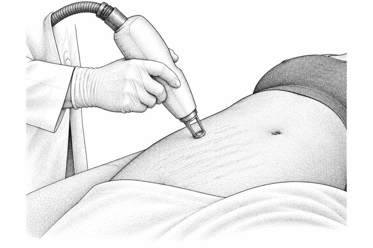
Laser treatment can make stretch marks flatter, smoother and closer to your natural skin tone, though rarely invisible. Here is what sessions, recovery and results usually look like.
What to expect from laser for stretch marks
A stretch mark can become flatter, smoother, and closer in color to surrounding skin after laser treatment, but it rarely disappears completely. Lasering stretch marks usually involves a series of treatments, often three or more sessions spaced about 3 to 4 weeks apart, with the laser choice based on the mark's color, age, location, and skin tone.
Stretch marks form when skin stretches faster than its collagen and elastic fibers can adapt. Newer marks may look pink, red, purple, or dark, while older striae albae are pale, white, and often more difficult to improve. Lasers create controlled heat in targeted areas to encourage collagen remodeling without treating all of the surrounding skin.
Non-ablative lasers usually cause mild redness for a day or two and have little downtime. Ablative fractional CO2 or Er:YAG lasers can produce more visible texture improvement, but commonly involve several days of redness, swelling, peeling, or fine crusting. Results build gradually over weeks, with collagen changes continuing for several months after the final session.
A consultation, skin-type assessment, and test spot are worth doing before treatment, especially for medium to dark skin tones that can develop post-inflammatory hyperpigmentation. Reviewing clinical stretch mark treatment options helps determine candidacy before scheduling a procedure.
Seek prompt medical care after treatment if blisters, increasing pain, spreading redness, drainage, or fever develop.
Which lasers are used for stretch marks
Striae distensae represent dermal scars resulting from mechanical stretching and hormonal fluctuations. During rapid growth, pregnancy, or weight changes, the structural integrity of the extracellular matrix breaks down. The dermis experiences micro-tearing, elastic fiber fragmentation, and disorganized collagen architecture. Lasers target these structural defects by delivering controlled thermal energy into the dermis. This process triggers neocollagenesis and tissue remodeling while sparing zones of healthy skin to accelerate recovery, a concept central to the laser scar reduction guide.
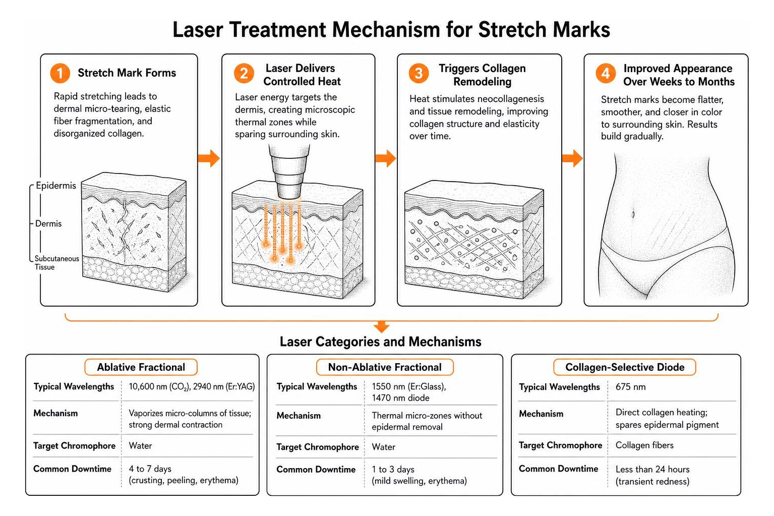
| Laser Category | Typical Wavelengths | Mechanism | Target Chromophore | Common Downtime |
|---|---|---|---|---|
| Ablative Fractional | 10,600 nm (CO2), 2940 nm (Er:YAG) | Vaporizes micro-columns of tissue; strong dermal contraction | Water | 4 to 7 days (crusting, peeling, erythema) |
| Non-Ablative Fractional | 1550 nm (Er:Glass), 1470 nm diode | Thermal micro-zones without epidermal removal | Water | 1 to 3 days (mild swelling, erythema) |
| Collagen-Selective Diode | 675 nm | Direct collagen heating; spares epidermal pigment | Collagen fibers | Less than 24 hours (transient redness) |
| Vascular / Deep Penetrating | 1064 nm (Nd:YAG) | Deep heating; targets microvasculature and deep dermis | Hemoglobin, deep water | 1 to 2 days (mild redness, warmth) |
| Photobiomodulation (Cold Laser) | 808 nm | Non-thermal cellular stimulation; stimulates ATP synthesis | Cytochrome c oxidase | None |
Ablative vs. non-ablative lasers
Ablative lasers remove microscopic columns of the epidermis while heating the underlying dermis. The fractional carbon dioxide (CO2) laser, operating at 10,600 nm, and the 2940 nm Erbium:YAG (Er:YAG) laser represent the two primary ablative systems. Because water is their primary target, these devices vaporize damaged scar tissue. This vaporization triggers a wound-healing cascade that thickens the epidermis and reorganizes dermal collagen fibers.
A randomized split-study evaluated 24 Asian adults comparing non-ablative fractional 1550 nm Er:Glass laser with ablative CO2 fractional laser for striae distensae. In this trial, both modalities delivered significant improvements. Skin elasticity increased from 59.05 to 69.55 with non-ablative laser and from 56.32 to 68.23 with fractional CO2 laser. Furthermore, the width of the widest stretch marks decreased from 7.00 mm to 4.55 mm in the non-ablative group and from 6.77 mm to 4.43 mm in the ablative group. While ablative CO2 lasers generated higher overall patient satisfaction (90.9% reported improvement compared to 81.8% in the non-ablative group), they also caused a significantly higher incidence of post-inflammatory hyperpigmentation (81.8% versus 36.4%) and longer downtime.
Non-ablative fractional lasers heat columns of the dermis to stimulate collagen production while leaving the stratum corneum intact. This preserves the skin's protective barrier, resulting in fewer post-procedure risks, less discomfort, and faster healing times.
Lasers for redness and pigment
Specialized wavelengths provide precise targeting when addressing specific components of stretch marks, such as color and localized collagen loss.
- 675 nm Diode Laser: This wavelength targets dermal collagen fibers directly instead of relying primarily on water absorption. In a clinical trial involving 32 female participants with striae distensae, three monthly sessions with a 675 nm laser reduced total Manchester Scar Scale scores from 14.16 (±1.30) at baseline to 10.06 (±1.32) at the 6-month follow-up. Color scores improved from 3.44 to 2.50, and texture scores improved from 2.00 to 1.37, with mild redness resolving within 24 hours as the only observed side effect.
- 1064 nm Long-Pulsed Nd:YAG: This wavelength penetrates deeply into tissue, making it suitable for stimulating deep dermal fibroblasts and addressing vascular redness in early stretch marks (striae rubrae). A comparative trial on 30 female patients with mature striae alba showed that three sessions of long-pulsed Nd:YAG laser reduced lesion length from 2.0±0.9 mm to 0.6±0.8 mm and lesion width from 1.4±0.5 mm to 0.5±0.5 mm.
- 1470 nm Non-Ablative Diode: Selective for dermal water, this wavelength creates controlled thermal coagulation zones. A pilot study of 20 female volunteers demonstrated that three sessions of 1470 nm laser reduced stretch mark depth by 47.06% (from 0.34 mm to 0.18 mm) and width by 33.13% (from 6.58 mm to 4.40 mm) on objective high-frequency ultrasound.
Light therapy for stretch marks
Photobiomodulation, also known as low-level light therapy (LLLT), uses non-thermal wavelengths (commonly 808 nm) to stimulate cellular activity. Instead of creating thermal injury, the light energy is absorbed by cytochrome c oxidase within cellular mitochondria. This absorption enhances adenosine triphosphate (ATP) synthesis, reduces inflammatory signaling, and supports fibroblast activity.
A randomized controlled trial on 30 patients with striae alba in Fitzpatrick skin phototypes III–IV compared 808 nm diode LLLT against fractional CO2 laser. The fractional CO2 laser achieved marked to near-total improvement in 80% of treated sites, while LLLT achieved marked improvement in 30% of sites. Combining both modalities produced marked improvement in only 10% of cases, though it slightly reduced the duration of side effects from the CO2 laser. LLLT produced zero adverse events, zero pain, and zero downtime, positioning it as an option for individuals seeking mild remodeling without recovery periods.
What a laser session involves
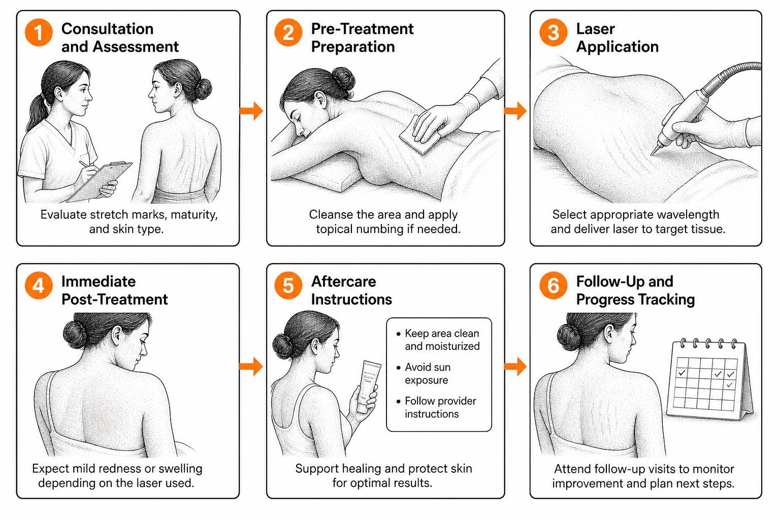
Receiving laser therapy for stretch marks follows a structured clinical protocol designed to ensure patient comfort, target tissue accurately, and minimize side effects.
How to prepare for treatment
The initial consultation involves determining the maturity of the stretch marks and evaluating the patient's Fitzpatrick skin phototype. Mature white marks (striae albae) require different energy settings than early red marks (striae rubrae).
- Medical History Review: The clinician screens for history of hypertrophic scarring, keloids, active infections, photosensitizing medications, and pregnancy.
- Skin Preparation: Patients must avoid direct sun exposure, tanning beds, and self-tanners for at least four weeks prior to treatment. Active skincare ingredients such as topical retinoids, glycolic acid, and salicylic acid should be discontinued 5 to 7 days before the session.
- Test Spotting: In darker skin types (Fitzpatrick III–VI), a test spot is placed in an inconspicuous area several days before full treatment to check for pigmentary changes or excessive thermal reactivity.
What happens during the laser treatment
During the active procedure, the practitioner follows specific steps to deliver the energy safely:
- Cleansing and Anesthesia: The treatment area is cleansed with an antiseptic solution. For ablative or deeper non-ablative treatments, a topical anesthetic cream (such as 4% lidocaine or compounded lidocaine/tetracaine) is applied for 45 to 60 minutes.
- Surface Cooling: To protect the epidermis and maintain comfort, clinicians employ integrated contact cooling tips, chilled conductive gels, or forced cold-air systems throughout the passes.
- Laser Passes: The provider guides the laser handpiece across the stretch marks. Standard non-ablative protocols often utilize 1 to 3 passes, whereas fractional ablative protocols may use a single, carefully calibrated pass with precise spot spacing (such as 0.8 to 1.5 mm).
- Sensory Experience: Patients typically feel a warm snapping or prickling sensation. Ablative treatments feel like deep heat accompanied by a mild sunburn sensation, with pain scores averaging around 6 out of 10 in clinical trials without nerve blocks. Non-ablative treatments are generally tolerated with lower discomfort scores (around 2 to 4 out of 10) during standard clinical laser treatment delivery.
Wound care and recovery after laser
Immediate post-treatment care focuses on soothing thermal heat and supporting barrier repair.
- Immediate Cooling: Chilled saline gauze or cold packs are placed over the treated zone for 15 to 20 minutes to reduce residual heat.
- Emollient Application: Occlusive or semi-occlusive soothing ointments (such as medical-grade petrolatum) are applied to hydrate the skin.
- Managing Normal Reactions: Redness (erythema) and mild swelling (edema) are expected. With non-ablative lasers, these resolve within 24 to 48 hours. With ablative lasers, microscopic crusting forms over 48 to 72 hours, resulting in a bronzed, sandpaper-like texture that naturally sloughs off over 5 to 7 days.
- Strict Sun Protection: Broad-spectrum mineral sunscreen (SPF 30 or higher) and protective clothing are mandatory to prevent ultraviolet rays from triggering post-inflammatory hyperpigmentation on healing skin.
Results and side effects by skin tone
Clinical outcomes are assessed using standardized tools, including the Manchester Scar Scale, the Global Aesthetic Improvement Scale (GAIS), and high-frequency ultrasound imaging to measure epidermal thickness and dermal collagen density.
Laser safety across Fitzpatrick skin types
Laser safety depends heavily on melanin concentration. In Fitzpatrick skin phototypes IV, V, and VI, epidermal melanin acts as a competing chromophore that can absorb laser energy unintentionally. When light intended for dermal water or collagen is absorbed by melanin, thermal injury occurs at the dermo-epidermal junction, increasing the risk of post-inflammatory hyperpigmentation (PIH) or hypopigmentation.
A study evaluating treatments for striae alba noted that fractional CO2 laser caused post-inflammatory hyperpigmentation in 16.7% of treated sites, and post-procedure erythema occurred in 83.3% of sites. In contrast, long-pulsed Nd:YAG laser produced erythema in only 13.3% of sites with 0% permanent PIH in the same patient population. For patients with darker complexions, non-ablative devices, long-pulsed Nd:YAG lasers, or conservative pulse energies combined with longer pulse durations provide a safer therapeutic window, consistent with protocols for laser scar removal for dark skin.
Common transient side effects across all skin tones include:
- Mild-to-moderate erythema (lasting 1 to 5 days)
- Localized edema and pinpoint crusting
- Mild itching during the re-epithelialization phase
Rare complications include persistent hyperpigmentation, permanent loss of pigment (hypopigmentation), secondary bacterial infection, or hypertrophic scarring caused by excessive pulse overlap.
How often to treat and how fast skin changes
Achieving noticeable texture improvement requires a series of treatments. Dermal remodeling is a biological process that unfolds across several stages:
- Treatment Number: 3 to 6 sessions depending on the depth and age of the stretch marks.
- Session Intervals: Spaced 3 to 4 weeks apart to allow complete re-epithelialization and initial collagen synthesis between passes.
- Histological Changes: Histopathological analysis shows that laser resurfacing increases both epidermal thickness and dermal collagen bundle density. A review on striae distensae modalities observed that non-ablative fractional and fractional CO2 scar resurfacing generate statistically significant structural increases in elastin and collagen fibers that continue to mature up to six months post-treatment.
Common questions about laser for stretch marks
How long between laser sessions
Most clinical protocols require between 3 and 5 sessions to achieve meaningful structural remodeling. Initial smoothing can often be seen within 4 weeks after the second session, but maximum collagen remodeling occurs between 3 and 6 months following the final laser treatment.
Managing discomfort during treatment
Discomfort varies by laser modality and energy level. Non-ablative and diode lasers cause mild to moderate stinging, often compared to the snap of a small rubber band against warm skin. Ablative fractional CO2 lasers generate deeper heat and higher discomfort, which is managed using topical numbing creams applied for an hour before the session along with continuous contact or forced-air cooling.
Limits of treating older white stretch marks
Lasers cannot completely erase mature white stretch marks because the original elastic fiber network cannot be recreated. However, laser treatments can significantly reduce their depth, narrow their width, improve overall skin elasticity, and blend their surface texture so that they match surrounding healthy skin much more closely.
Conclusion
Lasering stretch marks offers a practical clinical path for reducing the depth, width, and visible contrast of striae distensae. Whether selecting ablative fractional lasers for significant structural remodeling, non-ablative wavelengths for minimal downtime, or collagen-selective diodes for targeted stimulation, treatment plans should be tailored to individual skin phototypes and lesion maturity.
A personalized evaluation helps determine the appropriate laser modality, energy settings, and expected timeline. Evaluating tissue within stretch mark assessment systems helps classify specific lesions prior to intervention.
This content is for informational purposes only and does not constitute medical advice. Consult a qualified healthcare professional for diagnosis and treatment.
Works Cited
- Sadeghinia A, Emadi SN, Jamshidi S, Aryanian Z, Hatami P. “Combination of short pulse and smooth modality of Er: YAG laser versus short pulse alone in treatment of striae distensae: a split-lesion study.” Lasers in Medical Science, 2025.