Do Doctors Cut Through the Same C-Section Scar?
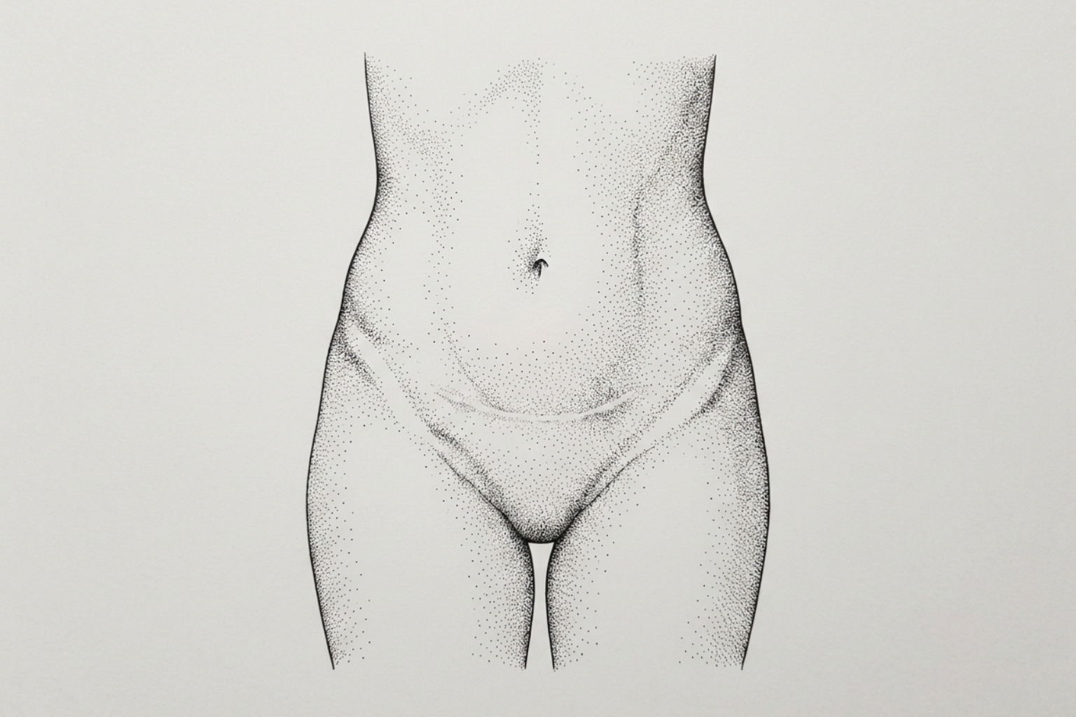
Why repeat C-sections often use the same incision
A prior C-section scar is usually reopened for a second or later cesarean delivery, most often through the same low, horizontal skin incision. This approach can avoid creating an additional visible scar, but the surgeon may adjust, extend, or remove part of the old scar when access, healing, or safety requires it.
The skin scar is only one layer of healing. Beneath it, prior surgery can leave internal scar tissue called adhesions, which may connect the uterus, bladder, bowel, or abdominal wall. Multiple C-section scars can make a repeat operation take longer, especially when adhesions are dense, but a flat, pale, or dark external scar does not reliably show what is happening inside.
Most skin healing takes about 4-6 weeks, while scar remodeling continues for several months. Gentle incision care, gradual movement after medical clearance, and realistic expectations can support recovery. More guidance is available in this C-section scar revision guide.
Contact the surgical or maternity care team promptly for fever, worsening pain, spreading redness, drainage, an opening incision, or foul-smelling discharge. Book a consultation if redness, itch, thickening, or tightness has not settled by two months.
The Scar Healing Editorial Team explains what repeat C-section incisions can mean for visible scars and the deeper tissues below them.
How surgeons re-enter the abdomen
During a repeat cesarean section, obstetricians make every effort to enter the abdomen through the original mark. Opening along the previous Pfannenstiel or low transverse incision avoids creating multiple parallel lines across the lower abdomen, which preserves aesthetic symmetry and reduces the overall surface area of cutaneous scarring.
When entering through an existing scar, the surgeon frequently performs a minor revision by excising the old, fibrotic skin margin. Removing the previous collagen band allows the fresh, well-vascularized edges of the skin to approximate cleanly, which often results in a neater cosmetic outcome than the first scar. However, if the prior scar healed with severe retraction, distortion, or significant widening, the surgeon may adjust the incision slightly to optimize surgical access, safety, and exposure. Reviewing available c-section scar revision options can help patients understand how cutaneous revisions are integrated into surgical planning.
Skin scar vs. uterine scar
The visible cutaneous line represents only the outermost layer of an abdominal delivery. An obstetrician must navigate multiple distinct tissue layers to reach the infant:
- Epidermis and dermis (external skin)
- Subcutaneous adipose tissue (superficial fat)
- Rectus sheath (fascial connective tissue)
- Rectus abdominis muscle separation
- Parietal peritoneum (abdominal cavity lining)
- Visceral peritoneum and vesicouterine fold
- Myometrium (muscular uterine wall)
- Amniotic sac and endometrium
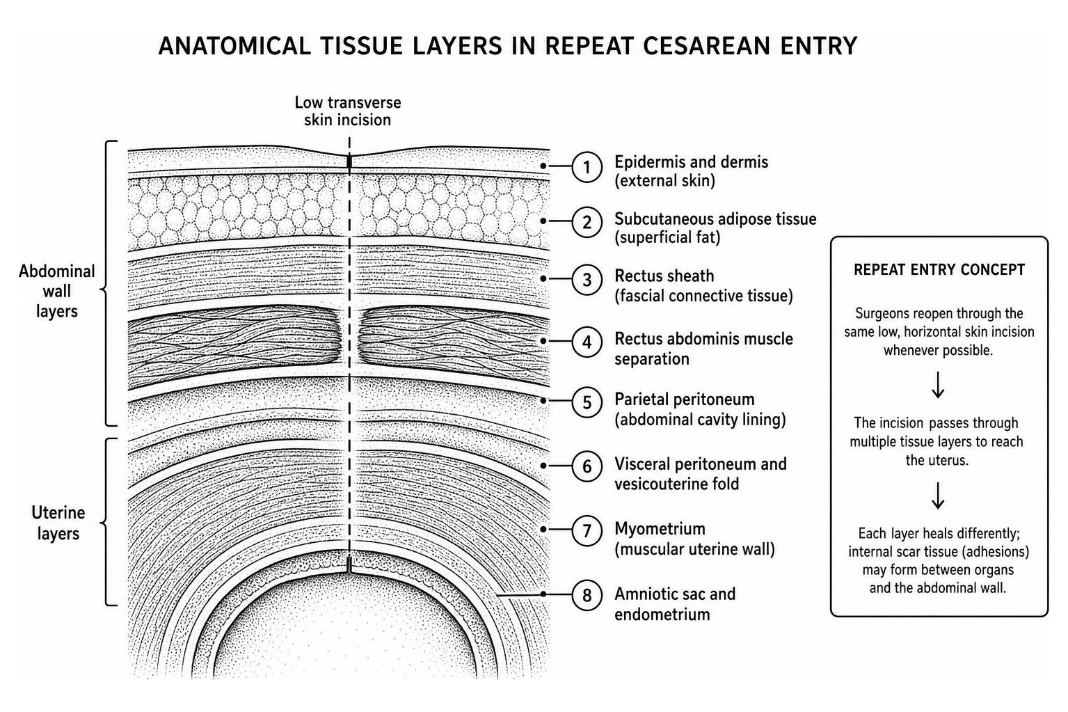
Healing varies across these individual strata. While the cutaneous layer may heal with minimal visible scarring, internal layers can form robust bands of connective tissue. The uterine incision, typically made in the lower uterine segment, heals with smooth muscle remodeling and collagen deposition. Understanding cesarean scar types helps differentiate between skin healing and the structural integrity of the underlying myometrium.
Is the old scar removed during surgery?
Uterine scar defects, commonly referred to as uterine niches or isthmoceles, can develop when the myometrial edges do not appose evenly or experience compromised perfusion during healing. To minimize this risk during subsequent deliveries, surgeons may perform an intentional resection of the old fibrous uterine scar before closing the new incision.
In a randomized controlled trial of 160 women published in BMC Pregnancy and Childbirth, researchers evaluated uterine scar resection during repeat delivery. The trial demonstrated that resecting the previous uterine scar reduced the incidence of uterine niche formation from 78.8% in the control group down to 45.0% in the resection group at 6 months postpartum. This surgical technique significantly improved residual myometrial thickness and decreased postoperative spotting days without increasing blood loss or total operative time.
Adhesions and other risks with multiple C-sections
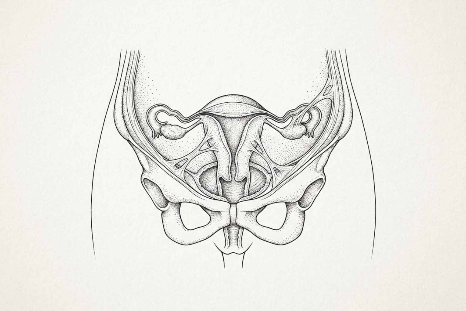
Every surgical entry into the peritoneal cavity causes localized inflammation, peritoneal drying, and microscopic tissue trauma. In response, the body deposits fibrin strands to seal injured surfaces. While these fibrin deposits normally dissolve through natural fibrinolysis, reduced local blood flow or repeated inflammation can cause these strands to mature into permanent fibrous bands known as intra-abdominal adhesions.
Adhesions can tether the anterior uterine wall to the posterior bladder base, anchor loops of small intestine to the abdominal wall, or fix the omentum across the pelvic brim. According to a prospective cohort study of 800 women published in Clinical and Experimental Obstetrics & Gynecology evaluating clinical research on adhesions in repeat cesareans, intra-abdominal adhesions were observed in 72.5% of patients undergoing repeat cesarean section. These internal bands complicate subsequent surgeries by distorting normal pelvic anatomy, extending the time required to deliver the infant safely, and elevating the risk of inadvertent bladder or bowel lacerations.
How bad adhesions can get
Adhesion density compounds with each successive abdominal delivery. Surgeons commonly grade adhesion severity using the Nair classification scale:
- Grade 0: No adhesions present.
- Grade 1: Filmy, thin adhesions easily separated by blunt dissection.
- Grade 2: Moderate adhesions with established vascularization requiring sharp dissection.
- Grade 3: Dense, cohesive adhesions between organs requiring extensive sharp dissection.
- Grade 4: Extensive adhesion bands creating an obliterated peritoneal cavity or "frozen pelvis."
In the 800-patient study published in Clinical and Experimental Obstetrics & Gynecology, 27.5% of women had no adhesions, 37.0% exhibited filmy adhesions, 17.0% had multiple dense bands, 15.5% had very dense adhesions, and 3.0% presented with a frozen pelvis. The authors noted that each additional previous cesarean section increased the odds of developing dense adhesions nearly threefold (odds ratio = 2.85, 95% confidence interval: 2.10–3.88).
A separate comparative study of 1,318 women published in the Pakistan Journal of Medical Sciences found that patients with four or more previous cesareans had an adhesion rate of 46.7%, compared to 23.8% in women with two to three prior deliveries. These dense bands contributed to significantly longer operating times (44.3 minutes versus 40.9 minutes), longer hospital stays (60.0 hours versus 52.2 hours), and higher blood transfusion rates. The risk of requiring a blood transfusion scaled progressively: 1.8% for one prior delivery, 2.6% for two, 4.3% for three, 4.6% for four, and 14.6% for five or more. When dense scar bands alter anatomical planes, surgeons must adapt their technique, a factor detailed in clinical reviews on laparoscopic considerations after multiple cesareans.
Does the skin scar predict adhesions inside?
A common clinical question is whether the visual presentation of an abdominal scar reflects the severity of internal adhesions. Many assume that a wide, pigmented, or raised scar indicates severe internal scarring, whereas a faint, thin scar suggests a clear abdominal cavity.
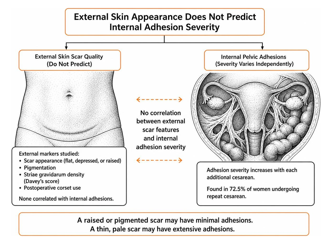
The prospective study of 800 women in Clinical and Experimental Obstetrics & Gynecology systematically evaluated external markers, including scar appearance (flat, depressed, or raised), pigmentation, striae gravidarum (stretch mark) density via Davey's score, and postoperative corset use. The researchers determined that none of these external features correlated with internal adhesion formation. A patient with severe striae and a raised external scar may have minimal intra-abdominal adhesions, while a patient with a thin, pale cutaneous scar may possess extensive pelvic adhesions. External skin healing reflects local dermal fibroblasts, whereas internal adhesions depend on peritoneal mesothelial cell healing and surgical trauma. Dermal risks can be explored further in clinical guides on body location risks for keloid and raised scars.
Long-Term Health Risks: Uterine Integrity, Isthmoceles, and Placenta Issues
The cumulative impact of multiple uterine incisions extends beyond surgical adhesions to include alterations in placental implantation and myometrial tensile strength. The table below outlines how specific clinical risks scale with repeated cesarean deliveries, based on obstetric literature summarized in guidance regarding evidence-based limits on repeat cesareans by the Mayo Clinic.
| Clinical Complication | Pathophysiological Mechanism | Risk Progression with Multiple Deliveries | Clinical Implication |
|---|---|---|---|
| Placenta Previa | Implantation over lower uterine segment scar tissue | Increases from ~0.3% in unscarred uterus to over 1-2% after multiple deliveries | Requires planned cesarean; increases antepartum hemorrhage risk |
| Placenta Accreta Spectrum (PAS) | Trophoblastic invasion into defective decidua and myometrium | Rises sharply; exceeds 40-50% in patients with previa and 3+ prior cesareans | Risk of severe hemorrhage, emergent hysterectomy, and ICU admission |
| Intra-Abdominal Adhesions | Incomplete peritoneal fibrinolysis and fibrous band maturation | Increases from 23.8% (2-3 cesareans) to 46.7%-54% (4-5+ cesareans) | Extended surgical time, elevated risk of bowel or bladder laceration |
| Blood Transfusion | Vascular disruption during dissection or abnormal placental separation | Progresses from 1.8% (1 prior CS) to 14.6% (5+ prior CS) | Heightened need for perioperative blood product preparation |
| Uterine Dehiscence / Rupture | Incomplete muscular remodeling of lower uterine segment scar | 0.5%–1% for single low transverse scar; increases to 2%–3% with multiple scars | Restricts trial of labor options; necessitates scheduled repeat delivery |
Treating isthmocele and C-section scar defects
An isthmocele, or cesarean scar defect, occurs when the myometrium at the incision site heals with a structural gap, creating an outpouching or reservoir along the anterior lower uterine segment. According to a clinical overview in the Journal of Minimally Invasive Gynecology, between 20% and 70% of women with at least one cesarean delivery demonstrate some degree of uterine niche on post-procedure ultrasound examination (Kremer et al., 2019).
Many isthmoceles remain asymptomatic, but larger defects can collect menstrual blood and inflammatory fluid, causing persistent postmenstrual brown spotting, chronic pelvic discomfort, dysmenorrhea, and secondary infertility. The trapped fluid within the niche can alter cervical mucus quality and disrupt normal endometrial receptivity.
Diagnosis is primarily achieved via transvaginal ultrasound or saline-infusion sonohysterography, ideally performed in the early follicular phase right after menstruation. On hysterosalpingography, scar defects manifest in varied configurations, ranging from thin linear clefts and wedge-shaped pockets to large saccular pseudodiverticula (Ahmadi et al., 2013). Management depends on the residual myometrial thickness and reproductive goals:
- Medical Management: Combined oral contraceptives or progestin therapy to suppress cyclic bleeding and alleviate spotting symptoms without correcting the anatomical pocket.
- Hysteroscopic Resection: Minimally invasive resectoscopic flattening of the distal and proximal niche edges, suitable for patients with adequate residual myometrium (typically greater than 3 mm).
- Laparoscopic / Robotic Excision: Complete surgical debridement of the fibrous niche margin followed by multilayered myometrial reconstruction, indicated when residual myometrium is thin (less than 2.5–3 mm) to restore uterine wall strength before future pregnancy.
Can you have a VBAC after multiple C-sections?
A Trial of Labor After Cesarean (TOLAC) leading to a Vaginal Birth After Cesarean (VBAC) is a safe alternative to scheduled surgery for many candidates. However, the presence of multiple uterine scars shifts the risk-benefit balance. According to clinical guidelines from the American College of Obstetricians and Gynecologists (ACOG), while a single low-transverse uterine scar carries a uterine rupture risk between 0.5% and 0.9%, the rupture risk rises to approximately 1% to 3% in individuals with two or more prior cesareans.
Obstetric guidelines evaluate several core factors when assessing VBAC candidacy after multiple deliveries, as outlined in clinical resources for evaluating pregnancy safety after multiple cesareans:
- Uterine Incision Direction: A documented low transverse incision carries the lowest rupture risk. Classical (vertical) incisions in the active, muscular upper segment are absolute contraindications for TOLAC due to a rupture rate approaching 10% based on ACOG guidelines.
- Prior Vaginal Delivery: ACOG reports that patients who have achieved at least one successful vaginal delivery (either before or after a cesarean) demonstrate VBAC success rates exceeding 80%.
- Interpregnancy Spacing: A spacing interval of less than 18 months between deliveries is associated with a two- to three-fold higher rate of uterine rupture, as myometrial collagen fibers require adequate time to regain baseline tensile stability.
- Institutional Capabilities: Safe labor trials require an obstetrical facility equipped for continuous electronic fetal monitoring, immediate surgical intervention, and emergency neonatal resuscitation.
Recovery timeline and treatment options
Recovery after a second or third cesarean delivery requires managing surgical pain alongside caring for an expanding family. The initial inflammatory phase transitions into tissue proliferation over the first three weeks, followed by deep collagen remodeling that continues for 12 to 18 months.
Managing recovery involves adhering to core postoperative milestones:
- Weeks 1–2: Focus on pain management, pelvic rest, wound hygiene, and gentle mobilization to prevent deep vein thrombosis.
- Weeks 3–6: Gradual resumption of routine non-strenuous daily activities; core strain and lifting objects heavier than the infant should be avoided.
- Weeks 6+: Formal obstetric evaluation for fascial integrity and clearance for targeted abdominal exercises, pelvic floor physical therapy, and topical scar remodeling.
Postoperative Wound Care and Early Healing
Proper early wound management establishes the foundation for clean scar maturation. The incision site should be kept clean, dry, and protected from unnecessary friction or tension during the early inflammatory phase. Patients should follow evidence-based protocols detailed in routine post-surgery incision care.
Washing the area gently with mild soap and water without direct scrubbing, patting the skin completely dry with a clean towel, and wearing loose, breathable clothing prevents maceration along the lower abdominal fold. Knowing when to begin postoperative scar treatment is critical: active topical interventions should only commence once the incision is fully re-epithelialized with no scabbing, drainage, or open gaps, which typically occurs between 2 and 4 weeks postpartum.
Non-invasive and in-office scar treatments
Once the skin barrier is fully intact, specialized modalities can help soften tissue stiffness, minimize hypertrophic thickening, and improve skin mobility across the lower abdomen.
- Medical-Grade Silicone Gel and Sheeting: Considered the primary conservative approach for surgical scars. Applying medical silicone sheets creates an occlusive, hydrating barrier that normalizes stratum corneum hydration and downregulates excessive fibroblast collagen production. Guidance on application protocols can be found under silicone treatments for scar management.
- Manual Scar Tissue Massage: Once cleared by an obstetrician (typically at 6–8 weeks), gentle manual mobilization across and parallel to the scar line helps disrupt cross-linked adhesions between the dermis, subcutaneous adipose tissue, and abdominal fascia. Reviewing scar tissue massage therapy protocols provides safe mechanical techniques for home care.
- Fractional Laser Therapy: For mature, thickened, or persistent red scars, dermatological energy devices such as pulsed-dye lasers (PDL) or non-ablative fractional lasers stimulate controlled microthermal remodeling. Clinical indications and expectations are detailed in the fractional laser scar reduction methods guide.
What to know about repeat C-sections
How many C-sections are safe?
Medical guidelines do not establish a fixed numeric maximum for cesarean deliveries. Every surgical history is unique, and some patients safely undergo four, five, or more procedures without major complications (Biler et al., 2017). However, cumulative risks such as dense adhesions, placenta accreta spectrum disorders, and surgical organ injury scale upward with each subsequent entry. The safety of additional deliveries depends on operative findings documented in prior surgical records, including the severity of internal scarring and lower uterine segment thickness.
Recovery and pain after repeat C-sections
Postoperative discomfort varies significantly among individuals. While the skin incision is often reopened cleanly, repeat surgeries can involve more intense uterine afterpains (involution cramping) as the multiparous uterus works harder to contract. If extensive adhesiolysis was required during the operation to mobilize the bladder or bowel, localized deep tissue soreness may feel more pronounced during the first 7 to 10 days of recovery.
How long to wait between pregnancies
Major obstetric organizations advise an interpregnancy interval of at least 18 to 24 months between a cesarean delivery and a subsequent conception. Spacing pregnancies allows the myometrial smooth muscle and collagen matrices within the lower uterine segment to regain structural integrity, which reduces the incidence of uterine scar dehiscence, placenta previa, and preterm birth in future pregnancies.
Conclusion
Navigating multiple cesarean deliveries involves understanding both external skin healing and deeper internal tissue remodeling. While surgeons typically reopen the original cutaneous incision to minimize new visible marks, the internal environment accumulates adhesions and myometrial changes that require thoughtful surgical planning and realistic recovery expectations. Open communication with an obstetrician regarding surgical history, prior operative notes, and personal family planning goals ensures the highest standard of maternal and neonatal safety. For an extensive overview of scar assessment and long-term dermal therapies, explore the comprehensive c-section scar guide.
This content is for informational purposes only and does not constitute medical advice. Consult a qualified healthcare professional for diagnosis and treatment.
Works Cited
Biler A, Ekin A, Ozcan A, Inan AH, Vural T, Toz E. "Is it safe to have multiple repeat cesarean sections? A high volume tertiary care center experience.." Pakistan journal of medical sciences, 2017. PMCID PMC5673710. Yusuf Ziya Kizildemir, Hacer Uyanikoglu, Ömer Tammo, Mehmet İncebiyik, Bekir Kahveci. "Predicting Intra-Abdominal Adhesions in Repeat Cesarean Sections: Scar Tissue Characteristics, Striae Gravidarum, and Corset Use." Clinical and Experimental Obstetrics & Gynecology, 2025. DOI 10.31083/ceog40126. Kremer TG, Ghiorzi IB, Dibi RP. "Isthmocele: an overview of diagnosis and treatment.." Revista da Associacao Medica Brasileira (1992), 2019. PMID 31166450. Ahmadi F, Torbati L, Akhbari F, Shahrzad G. "Appearance of uterine scar due to previous cesarean section on hysterosalpingography: various shapes, locations and sizes.." Iranian journal of radiology : a quarterly journal published by the Iranian Radiological Society, 2013. PMCID PMC3767017.