4 Ways Doctors Treat and Remove Contracture Scars
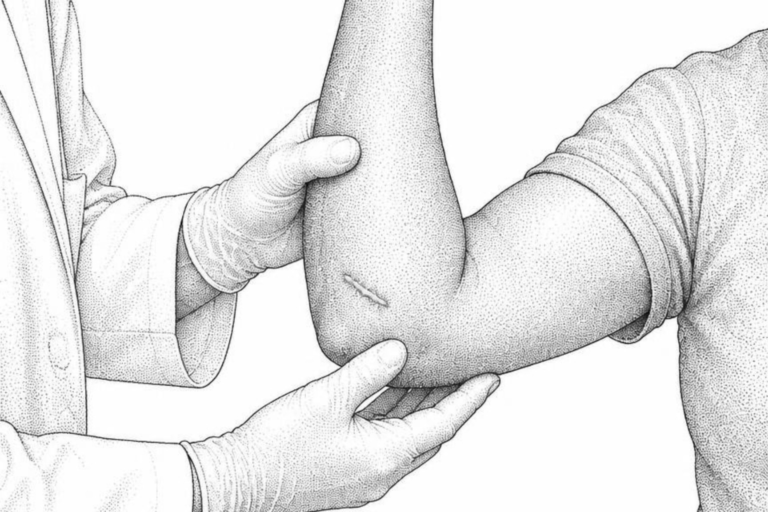
A contracture scar pulls skin tight across a joint and can limit movement. Doctors combine physical therapy, laser, injections and surgical release. Here is how each option works.
Treating contractures that limit movement
Tight fibrotic bands pulling across a joint restrict functional mobility and exert continuous mechanical tension on surrounding skin. Clinicians improve functional range by combining four modalities: physical therapy and splinting, laser treatment, intralesional pharmacotherapy for raised or thick tissue, and surgical release to rearrange the skin when movement remains restricted. Patients who want to know how to remove contracture scars should understand that the goal is functional improvement, not complete removal.
The scar cannot be eliminated completely, but medical intervention can make it softer, flatter, less tight, and more functional. Early care matters because scars often keep changing for 6 to 12 months; stretching, guided motion, massage after full wound closure, silicone, and pressure therapy may help preserve range of motion while tissue matures. A mature contracture may require surgical release, such as Z-plasty, a skin graft, or a local flap, as detailed in clinical resources on contracture scars. Patients should schedule a clinical consultation if the scar prevents full joint extension, tightens progressively, or remains raised, pruritic, or indurated after two months, while signs of acute infection require prompt medical evaluation.
The Scar Healing Editorial Team outlines the clinical mechanisms, procedural pathways, and recovery expectations across these therapeutic options.
How contracture scars are treated
Contracture scars develop when an extensive injury, deep dermal burn, or surgical wound heals by contracting the surrounding tissue. During normal wound repair, specialized cells called myofibroblasts contract to pull the wound margins together. In extensive injuries, these cells produce excessive extracellular matrix components and persistent tension. This mechanical force leads to rigid structural bands that shorten the skin surface.
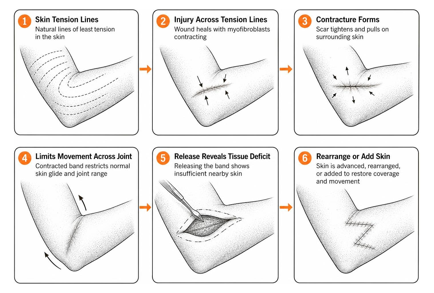
When a scar contracts across a joint, it creates a true tissue deficit. Releasing the contracted band reveals that the native skin area is insufficient to cover the underlying structures. The Textbook on Scar Management chapter on contractures notes that contractures frequently impact regions with high mobility, including the neck, axilla, and digits.
Understanding contracture scar management begins by recognizing that complete physical removal is not surgically possible without creating a secondary surgical site. Medical care focuses on eliminating the tension band, replenishing missing surface area, and restoring normal joint kinematics. A comprehensive overview of contracture scars explains the full range of treatment options.
Surgeons categorize the management pathway into active remodeling and mature stabilization stages. Surgical intervention is typically deferred for 6 to 12 months post-injury to allow natural remodeling to conclude. Immediate non-surgical therapies manage tissue stiffness during this intermediate window.
Surgical release and skin rearrangement
When conservative measures fail to preserve joint motion, surgical release becomes necessary. The operative approach involves incising the restrictive scar band, excising deep fibrotic cords down to healthy subcutaneous tissue, and redistributing mechanical tension along relaxed skin tension lines. Reconstructive surgeons evaluate anatomical depth, regional vascular supply, and adjacent skin elasticity to plan the appropriate reconstruction, as outlined in the StatPearls review on scar revision.
How Z-plasty works
Z-plasty is a foundational local transposition flap technique used to disrupt linear contracture bands. The surgeon creates a central incision along the scar line flanked by two equal-length limbs angled at approximately 60 degrees, forming a classic "Z" configuration.
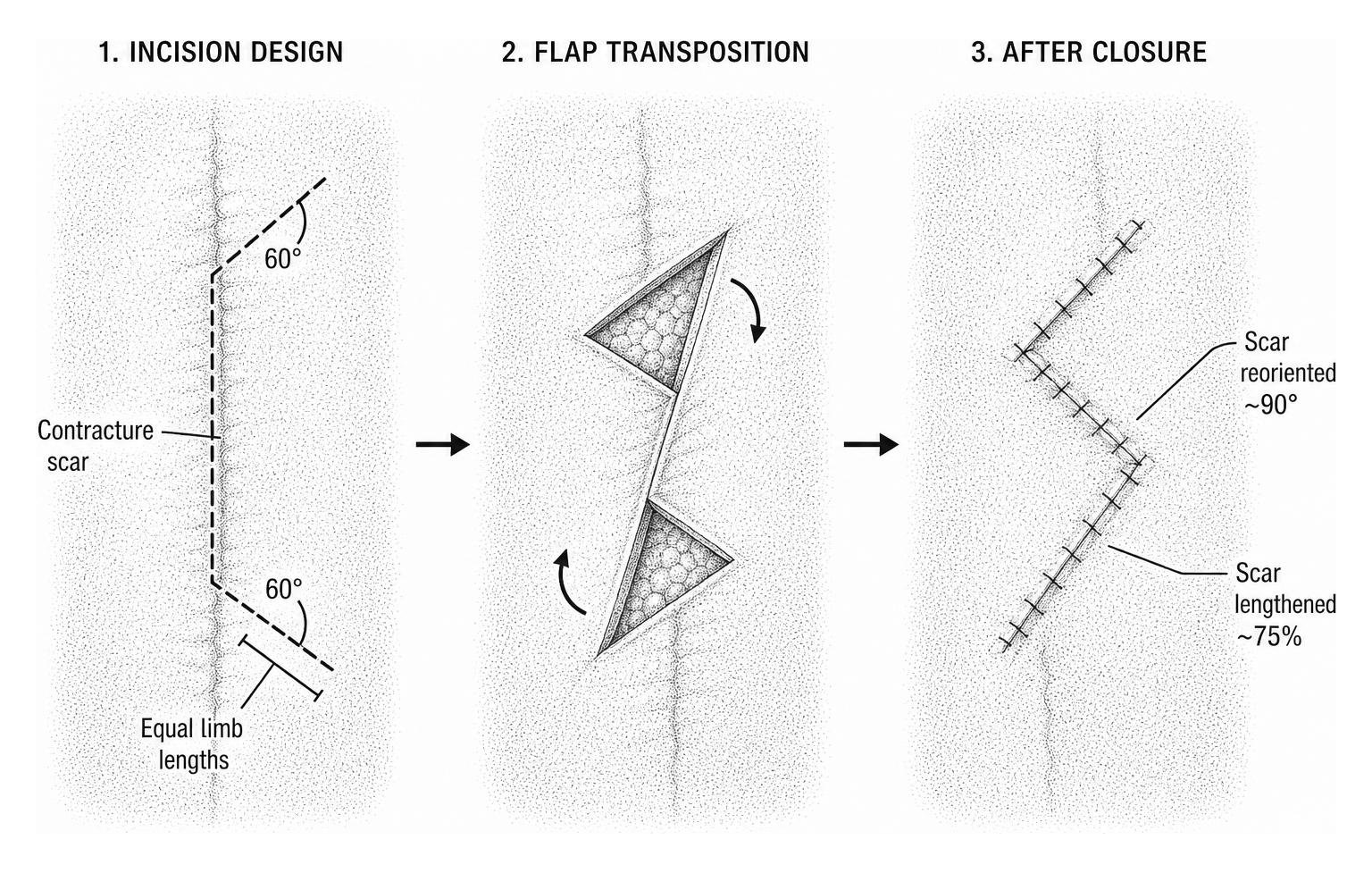
Transposing these triangular flaps accomplishes two mechanical goals:
- It reorients the scar vector by approximately 90 degrees, aligning it with natural skin folds.
- It lengthens the original scar line by approximately 75 percent based on standard geometric transposition models (a 1.7-fold extension factor relative to limb length).
In delicate structures such as the digits, limb lengths are calibrated between 5 and 8 mm, while hand and web-space releases accommodate 10 to 15 mm incisions. For lengthy linear bands, surgeons employ multiple continuous or running Z-plasties to redistribute tension uniformly across the joint axis, providing targeted surgical scar revision.
Skin grafts and flap surgery
Extensive planar contractures lack pliable adjacent tissue, precluding local transposition flaps. In these scenarios, surgeons utilize full-thickness skin grafts, local advancement flaps, or pedicled perforator flaps.
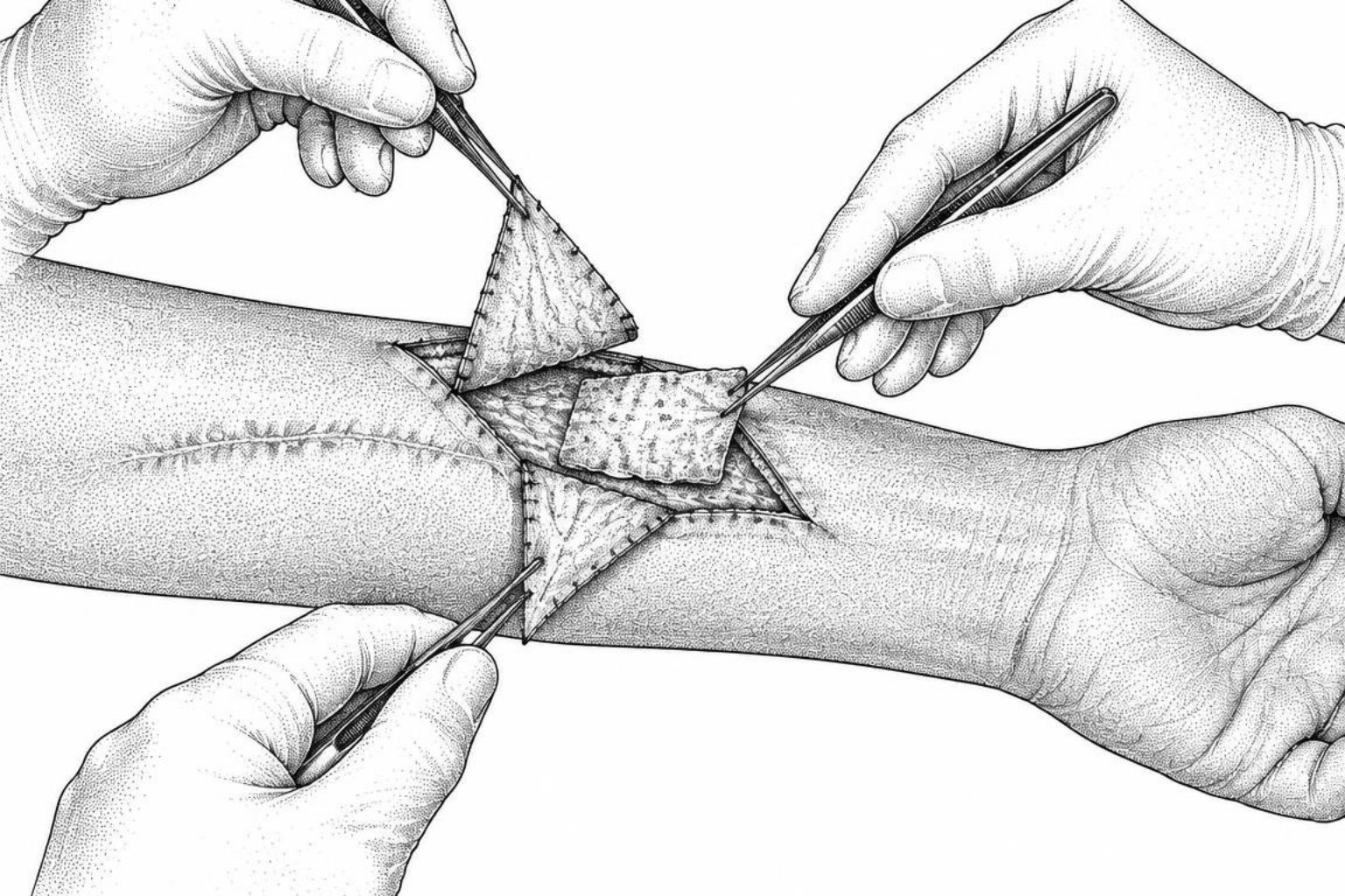
A clinical strategy published in the Journal of Clinical Medicine on hand scar contractures notes that thin split-thickness grafts frequently undergo secondary contraction, making full-thickness skin grafts or vascularized flaps preferable for functional areas. Full-thickness grafts include the complete dermis, which resists myofibroblast-driven shrinkage and matches regional skin characteristics.
For complex three-dimensional defects with exposed tendon or bone, regional pedicled perforator flaps or microvascular free tissue transfers bring their own independent blood supply. This robust vascularization ensures durable defect coverage and minimizes recurrent tension bands, which is essential for long-term burn scar management.
How fractional lasers loosen contracture scars
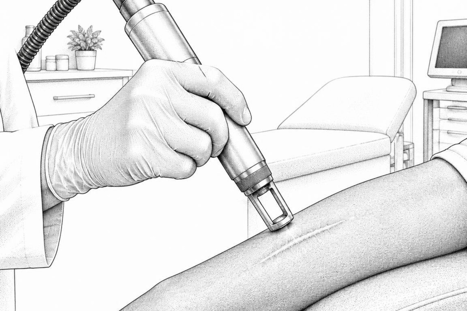
Laser modalities provide non-surgical options to soften rigid scar tissue and improve compliance. Fractional ablative lasers create precise columns of thermal micro-injury that stimulate normal collagen remodeling without removing the entire epidermal surface.
CO2 laser for contracture scars
Fractional carbon dioxide (CO2) lasers emitting at a 10,600 nm wavelength penetrate deep into the fibrotic dermis. The concentrated microthermal zones vaporize dense, disorganized type I collagen bundles. This controlled thermal coagulation triggers heat-shock protein expression and activates matrix metalloproteinases, replacing rigid scar tissue with organized type III collagen and functional elastin.
Clinical protocols for laser scar reduction typically involve multiple treatment sessions spaced 6 to 10 weeks apart:
- Energy densities and pulse durations are adjusted according to scar thickness and tissue depth.
- Conservative fluence levels with spot test areas are implemented for darker skin phototypes to reduce post-inflammatory pigment alterations.
- Topical or intralesional anti-inflammatory agents may be delivered immediately following ablation through laser-assisted drug delivery, facilitating deep dermal penetration.
Steroid injections to soften scars
Pharmacologic injections alter the cellular machinery within active, raised contracture bands. Intralesional corticosteroids, primarily triamcinolone acetonide (typically 10 to 40 mg/mL), suppress fibroblast proliferation, diminish pro-inflammatory cytokine secretion, and inhibit collagen synthesis.
- Triamcinolone Acetonide: Softens dense structural bands, reduces localized pruritus, and flattens elevated hypertrophic margins.
- 5-Fluorouracil (5-FU): An antimetabolite frequently combined with corticosteroids to arrest pyrimidine synthesis in proliferating fibroblasts, reducing collagen generation while lowering corticosteroid-associated skin atrophy.
- Collagenase Injections: Enzymatic preparations that cleave triple-helical collagen fibers, facilitating mechanical elongation in targeted fibrotic cords.
These agents are injected directly into the dense core of the scar instead of healthy subcutaneous fat, preventing adjacent dermal thinning, hypopigmentation, or telangiectasia.
Splints, compression and physical therapy
Mechanical force guides biological tissue remodeling. Physical therapy and targeted immobilization prevent recurrent scar contracture during active maturation and following surgical intervention.
Postoperative and non-surgical rehabilitation relies on static, dynamic, and progressive orthotic devices:
- Static Splints: Maintain the affected joint in maximum elongated extension during periods of immobility, particularly overnight.
- Dynamic Splints: Apply low-load, prolonged mechanical stretch using elastic components, encouraging cellular realignment without tearing remodeling tissues.
- Pressure Garments: Provide 15 to 30 mmHg of continuous surface pressure for up to 23 hours daily, reducing local tissue edema, limiting local blood flow, and suppressing excessive extracellular matrix synthesis.
Following surgical contracture release, nighttime splinting is maintained for at least three months to counteract secondary tissue shrinkage.
Manual massage and stretching
Manual physical therapy techniques modify cross-linked collagen networks once surgical incisions or traumatic wounds achieve complete epithelial closure. A 2025 systemic review in PLOS One involving non-surgical interventions recognized massage therapy as an effective approach for improving scar pliability, vascularity, and thickness.
Therapists apply myofascial release, transverse friction massage, and targeted scar tissue massage to break down deep dermal adhesions binding the skin to underlying fascial sheaths. Regular, structured tension application promotes structural fiber realignment parallel to the mechanical stress vectors, restoring normal gliding planes and tissue flexibility.
Common questions about removing contracture scars
Can a contracture scar be fully removed?
No medical or surgical procedure can completely eliminate a contracture scar or restore the skin to its original, uninjured state. Surgical interventions replace a tight, functionally restrictive scar with a planned, tension-free surgical closure or graft. For patients researching how to remove contracture scars, the realistic objective is restoring functional range of motion, reducing local discomfort, and improving tissue softness and appearance.
Recovery timeline after surgery
Surgical incisions typically achieve preliminary wound closure within 10 to 14 days. However, complete recovery and scar maturation require 6 to 18 months. During this period, the healing tissue gradually remodels, becoming softer, flatter, and paler. Patients generally maintain nighttime splinting for at least three months and participate in regular physical therapy throughout the first year to preserve motion.
When to start treatment
Non-surgical interventions, including passive stretching, custom splinting, and silicone application, should begin as soon as the wound attains full epithelialization. Elective surgical contracture release is generally performed after the scar has completed active remodeling, typically 6 to 12 months after injury. Surgery may be performed earlier if severe contractures cause joint subluxation, expose critical neurovascular structures, or present acute functional impairment.
Key takeaways
Managing a contracture scar requires a comprehensive approach tailored to the scar's maturity, anatomical location, and functional impact. By combining surgical revision, advanced laser resurfacing, targeted intralesional pharmacotherapy, and rigorous physical rehabilitation, patients can overcome mechanical restrictions and restore joint mobility. Individuals seeking a structured evaluation can take the next step with a personalized scar assessment to determine the appropriate treatment pathway.
Works Cited
- Cho H, Ono S, Chung KC. “Management of Scar Contractures of the Hand-Our Therapeutic Strategy and Challenges.” Journal of clinical medicine, 2024.
- Masanovic MG, Téot L. “Scar Contractures.” Textbook on Scar Management (Téot L, Mustoe TA, Middelkoop E, et al., eds.), Springer, 2020.
- Tullington JE, Gemma R. “Scar Revision.” StatPearls Publishing, 2023.
- Dastagir K, Obed D, Bucher F, Hofmann T, Koyro KI, Vogt PM. “Non-Invasive and Surgical Modalities for Scar Management: A Clinical Algorithm.” Journal of personalized medicine, 2021.
- Raborn LN, Janis JE. “Prevention and Treatment of Burn Scar Contracture: A Practical Review.” Plastic and Reconstructive Surgery - Global Open, 2024.
This content is for informational purposes only and does not constitute medical advice. Consult a qualified healthcare professional for diagnosis and treatment.