Why Your Skin Can't Keep Up: The Science of Fibroblast Dysfunction in Diabetes
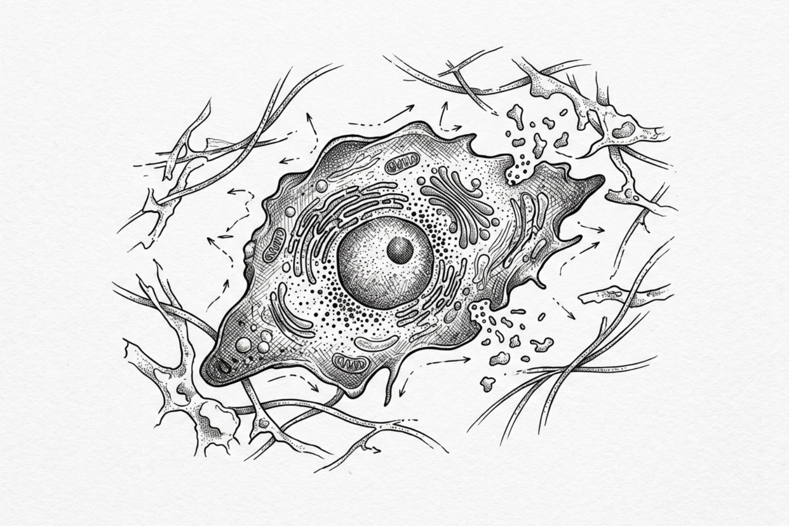
In diabetes, the skin's repair cells lose the ability to move, sense oxygen, and rebuild tissue. Here's the science behind slow-healing wounds and diabetic foot ulcers — and the therapies aiming to reverse it.
How Diabetes Disrupts the Body's Wound-Repair Process
Fibroblast dysfunction diabetes describes a well-documented pattern of cellular failure in which the skin's primary repair cells — fibroblasts — lose their ability to function normally under diabetic conditions, particularly in the presence of chronic high blood sugar (hyperglycemia).
Key impairments observed in diabetic fibroblasts include:
- Severely reduced migration — diabetic fibroblasts move up to 75% less than healthy fibroblasts, slowing wound closure
- Dramatically lower VEGF production — a sevenfold reduction in vascular endothelial growth factor (VEGF), a protein critical for new blood vessel formation
- Failed response to low oxygen (hypoxia) — healthy fibroblasts triple their VEGF output under low-oxygen conditions; diabetic fibroblasts show no such response
- Elevated tissue-degrading enzymes — diabetic fibroblasts produce roughly twice the level of pro-MMP-9, an enzyme that breaks down the structural scaffolding wounds need to close
- Impaired proliferation — associated with excess l-lactate accumulation driven by high glucose environments
Fibroblasts are the connective tissue cells responsible for depositing collagen, remodeling the extracellular matrix (ECM) — the structural framework of skin — and signaling for new blood vessel growth after injury. In healthy skin, they are the body's core repair crew, moving into wound sites, laying down new tissue, and coordinating healing across multiple phases.
In diabetes, that crew doesn't show up properly.
Research using diabetic animal models has shown that these impairments are not simply a genetic quirk. They appear after the onset of hyperglycemia — not before it. Fibroblasts from newborn diabetic mice function normally. It is the sustained exposure to high blood sugar that corrupts their behavior over time.
This has major real-world consequences. Diabetic foot ulcers (DFUs) — chronic, non-healing wounds that affect an estimated 40 to 60 million people worldwide — are a direct downstream result of this cellular breakdown. Up to 25% of people with Type 2 diabetes will develop a foot ulcer during their lifetime, and the annual cost to the US healthcare system alone is estimated at $9 to $13 billion.
Understanding why fibroblasts fail in diabetes is the first step toward understanding how these wounds can be better treated — and why standard wound care so often falls short.
Why fibroblasts fail in diabetes
The biological failure of the diabetic fibroblast is not a single defect but a cascade of mechanical and chemical impairments. When a wound occurs, the body relies on fibroblasts to sense the injury, migrate to the site, and release growth factors that stimulate the growth of new blood vessels (angiogenesis). In patients with diabetes, this sequence is fundamentally broken.
Why repair cells stop moving
One of the most striking observations in clinical research is the "lazy" behavior of these cells. Evidence indicates that adult diabetic mouse fibroblasts exhibit a 75% reduction in migration compared to normal fibroblasts (P < 0.001). In a healthy state, fibroblasts are highly mobile, crawling through the extracellular matrix to reach the wound "gap." In a diabetic environment, they remain largely stationary, failing to bridge the wound.
Weaker blood-vessel signals (VEGF)
A critical component of healing is the production of Vascular Endothelial Growth Factor (VEGF). This protein signals the body to build new capillaries to bring oxygen and nutrients to the healing tissue. Research published in the American Journal of Pathology found that adult diabetic fibroblasts exhibited a sevenfold impairment in VEGF production (4.5 ± 1.3 pg/ml versus 34.8 ± 3.3 pg/ml).
Furthermore, diabetic fibroblasts lose their ability to respond to environmental cues. In healthy tissue, low oxygen levels (hypoxia) act as a "flare gun," signaling cells to produce more VEGF. While wild-type fibroblast production of VEGF increases threefold in response to hypoxia, diabetic fibroblasts show no upregulation at all. This failure to respond to hypoxia is a primary reason why diabetic wounds remain ischemic (starved of blood flow).
Too much MMP-9 breaks down tissue
Healing requires a delicate balance between building new tissue and clearing away damaged proteins. This balance is managed by Matrix Metalloproteinases (MMPs). Diabetic fibroblasts have been shown to produce twice the amount of pro-matrix metalloproteinase-9 (pro-MMP-9) as normal fibroblasts. Excessive MMP-9 levels lead to the over-degradation of the extracellular matrix, effectively dissolving the "scaffolding" faster than the cells can build it. This creates a cycle of chronic inflammation and prevents the wound from transitioning out of the early inflammatory phase.
How high blood sugar harms fibroblasts
The primary driver of these cellular defects is chronic hyperglycemia. High glucose levels do not just sit in the blood; they alter the internal chemistry of the cells through several pathways.
- L-lactate Accumulation: Studies of fibroblasts derived from chronic diabetic wounds show significantly higher levels of l-lactate (6.3 ± 0.7 mmol/L) compared to non-diabetic wounds (2.1 ± 0.3 mmol/L). High levels of l-lactate have been shown to reduce DNA content in fibroblasts by up to 58%, directly inhibiting their ability to proliferate and repair tissue.
- Compromised DNA Repair: Chronic hyperglycemia triggers metabolic reprogramming that depletes cellular NAD+ pools. This depletion impairs the Non-Homologous End Joining (NHEJ) pathway, which is responsible for repairing DNA double-strand breaks. Research indicates that compromised DNA repair is a significant factor in diabetes-associated fibrosis. When DNA repair is compromised, fibroblasts enter a state of senescence (cellular aging) or undergo apoptosis (cell death).
- Reducing Sugars: It is not just glucose that causes damage. Other reducing sugars like ribose and fructose have been shown to have an even stronger negative impact on DNA repair efficiency than glucose, correlating with their higher reducing capacity and ability to induce oxidative stress.
Is the damage permanent?
A pivotal question in diabetic research is whether these fibroblast defects are hardwired (genetic) or acquired (environmental). Evidence from neonatal (pre-diabetic) db/db mouse models suggests the latter.
Fibroblasts harvested from newborn diabetic-prone mice — before they develop high blood sugar — function almost identically to healthy fibroblasts. They produce normal levels of VEGF and migrate effectively. This suggests that fibroblast dysfunction diabetes is an acquired condition resulting from prolonged exposure to a hyperglycemic environment. This is a crucial distinction because it suggests that the damage might be preventable or even reversible if the metabolic environment is corrected early enough.
Interestingly, the impairment is selective. While migration and VEGF production are severely hampered, some studies suggest that certain aspects of fibroblast function, such as basic proliferation and the development of a senescence-associated secretory phenotype (SASP), may persist, albeit in a maladaptive way that promotes chronic inflammation rather than healing, as explored in Frontiers in Immunology.
What this means for wounds and foot ulcers
The clinical manifestation of these cellular failures is most visible in the development of Diabetic Foot Ulcers (DFUs). These ulcers are the leading cause of hospitalization for diabetic patients, accounting for up to 25% of all admissions.
- Prevalence: Between 40 and 60 million people globally suffer from DFUs.
- Risk: Up to 25% of patients with Type 2 Diabetes Mellitus will develop an ulcer.
- Economic Burden: The annual cost of treating DFUs in the United States is estimated to be between $9 and $13 billion.
- Mortality: The stakes are high; the 5-year survival rate after a diabetes-related amputation is only about 50%, a figure comparable to many aggressive cancers.
The failure of the "repair crew" means these wounds often stay "stalled" for months or years. Without adequate migration to close the gap, or VEGF to provide blood flow, the wound becomes a breeding ground for infection. For more information on managing complex wounds, see our surgery scar treatment complete guide.
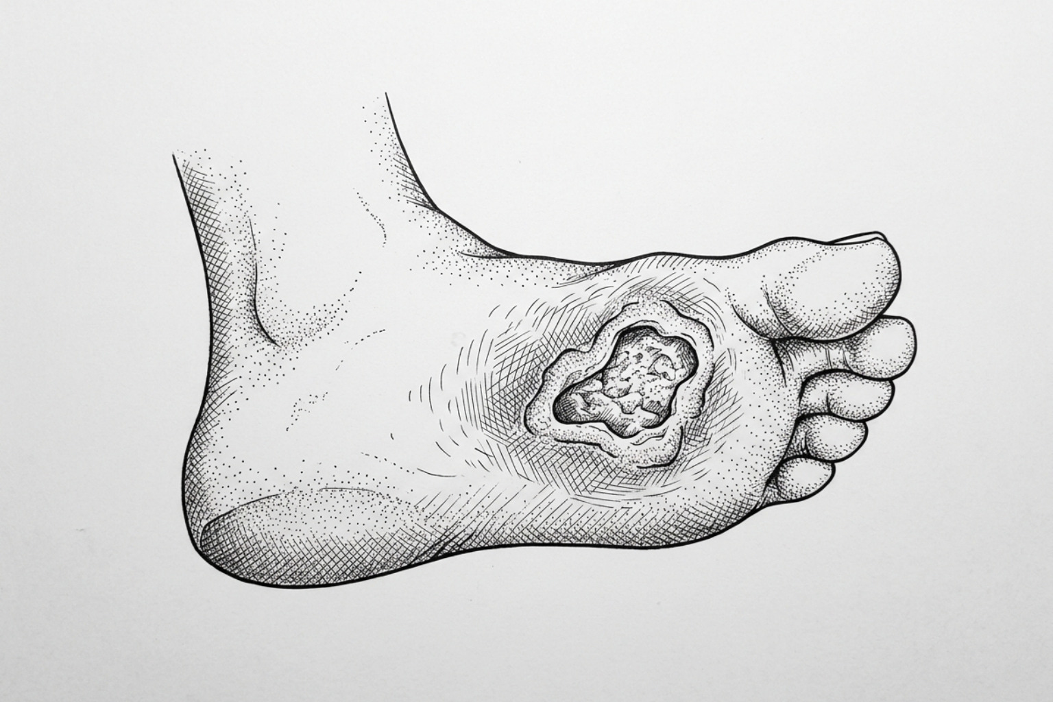
Different fibroblasts in foot ulcers
Recent advances in single-cell RNA sequencing (scRNA-seq) have revealed that not all fibroblasts are the same. Dermal fibroblasts are divided into two main populations:
- Papillary Fibroblasts: Located in the superficial dermis, these are spindle-shaped and are primarily involved in hair follicle regulation and re-epithelialization. They respond strongly to Wnt and Notch signaling pathways.
- Reticular Fibroblasts: Found in the deeper dermis, these are more stellate (star-shaped) and are responsible for producing the bulk of the collagen and extracellular matrix. They are driven by TGF-β signaling.
In diabetic wounds, this heterogeneity is disrupted. Research published in the Biomedicines has identified specific CD34+ fibroblast clusters that are downregulated in diabetic wounds compared to healthy healing tissue. Furthermore, fibroblasts in chronic diabetic environments can act as "immune sentinel cells," producing chemokines that recruit inflammatory cells but fail to resolve the inflammation, sometimes even forming tertiary lymphoid structures (TLS) that perpetuate a state of chronic wound "stagnation."
What happens at the molecular level
The "memory" of hyperglycemia in fibroblasts is often maintained through epigenetic modifications — changes to how genes are turned on or off without changing the DNA sequence itself.
The AGE/RAGE Axis
One of the most well-studied pathways involves Advanced Glycation End-products (AGEs). These are proteins or lipids that become glycated after exposure to high sugar. These AGEs bind to the Receptor for Advanced Glycation End-products (RAGE) on the fibroblast surface. This activation triggers a pro-inflammatory cascade involving the TGF-β/Smad pathway, leading to excessive but disorganized collagen deposition, which contributes to organ-specific fibrosis in the heart, kidneys, and skin.
Fibroblasts in the heart
In the heart, fibroblast dysfunction diabetes manifests as diabetic cardiomyopathy. High glucose activates the local renin-angiotensin system (RAS) within cardiac fibroblasts, increasing the production of collagen I and III. This leads to a stiffening of the heart tissue and diastolic dysfunction. Unlike skin fibroblasts, which fail to produce enough matrix to close a wound, cardiac fibroblasts in diabetes often produce too much matrix, leading to scarring and heart failure.
The "metabolic memory" effect
Even after blood sugar is brought under control, fibroblasts may continue to behave as if they are in a hyperglycemic state. This is due to DNA methylation and histone modifications that "lock" the cell into a dysfunctional state. Understanding these epigenetic locks is a major focus of current research into reversing diabetic complications.
How to restore fibroblast function
Addressing the "lazy fibroblast" requires moving beyond simple wound dressings. Current research is exploring several high-tech interventions:
| Functional Metric | Healthy Fibroblast | Diabetic Fibroblast | Potential Intervention |
|---|---|---|---|
| Migration | High (100%) | Low (25%) | Pathway modulation (Wnt/Notch) |
| VEGF Production | High (34.8 pg/ml) | Low (4.5 pg/ml) | VEGF supplementation/Gene therapy |
| Hypoxia Response | 3x Increase | No Change | RAGE inhibitors / Hypoxia mimetics |
| MMP-9 Levels | Balanced | 2x Elevation | MMP inhibitors / TIMP-1 restoration |
| DNA Repair | Efficient | Compromised | NAD+ precursors / RAGE phosphomimetics |
Emerging Treatments
- MMP Inhibition: Since diabetic fibroblasts overproduce matrix-degrading enzymes, using inhibitors to rebalance the MMP/TIMP ratio can help stabilize the wound environment.
- VEGF Supplementation: Directly applying growth factors or using viral vectors to "re-teach" fibroblasts to produce VEGF has shown promise in animal models.
- Targeting Macrophage-Fibroblast Crosstalk: In diabetic wounds, the communication between immune cells (macrophages) and fibroblasts is broken. Paracrine signaling from pro-inflammatory M1 macrophages further suppresses fibroblast migration. Immunomodulatory therapies that shift macrophages to a pro-healing M2 phenotype can help "wake up" the dormant fibroblasts.
- Restoring DNA Repair: Experimental treatments using nuclear phosphomimetic RAGE have successfully restored DNA repair capacity in diabetic models, reducing cellular senescence and fibrosis.
For those concerned about how these dysfunctions affect long-term scarring, resources like our surgical scar revision complete guide offer insights into corrective procedures.
Frequently Asked Questions
Why do diabetic wounds take so long to heal?
The primary reason is a combination of poor blood flow and fibroblast dysfunction diabetes. Because diabetic fibroblasts migrate 75% less and fail to produce the necessary VEGF to grow new blood vessels, the wound lacks the physical cellular presence and the nutrient supply required for closure. This is exacerbated by high levels of MMP-9, which break down new tissue as fast as it is formed.
Can high blood sugar be reversed to fix fibroblast function?
Strict glycemic control is the most effective way to prevent the onset of these dysfunctions. Research on neonatal models shows that the defects are acquired, not genetic. However, due to "metabolic memory" (epigenetic changes), simply lowering blood sugar after years of hyperglycemia may not immediately restore fibroblast function, often requiring additional therapeutic intervention.
What is the role of MMP-9 in diabetic ulcers?
MMP-9 is an enzyme that digests the extracellular matrix. In diabetic fibroblasts, pro-MMP-9 production is doubled. This creates an environment where the structural framework of the skin is constantly being degraded, preventing the wound from moving from the inflammatory phase to the proliferative (building) phase of healing.
Key Takeaways: Getting the Repair Crew Back to Work
The "lazy fibroblast" is not a choice made by the cell, but a consequence of a toxic metabolic environment. Chronic hyperglycemia re-programs these essential repair cells, stripping them of their ability to move, breathe, and build. This breakdown is the fundamental cause of the millions of diabetic foot ulcers that lead to suffering and amputation globally every year.
As we look toward 2026 and beyond, the focus of diabetic wound care is shifting from "covering the wound" to "re-activating the cell." By targeting specific molecular pathways like the AGE/RAGE axis and restoring DNA repair mechanisms, we may finally be able to get the body's repair crew back to work. If you are monitoring a healing wound, you can use our scar assessment tool to track your progress.
Works Cited
- Lerman, O. Z., et al. "Cellular Dysfunction in the Diabetic Fibroblast: Impairment in Migration, Vascular Endothelial Growth Factor Production, and Response to Hypoxia." American Journal of Pathology, 2003.
- Liu, Y., et al. "Fibroblasts: Immunomodulatory Factors in Refractory Diabetic Wound Healing." Frontiers in Immunology, 2022.
- Kumar, V., et al. "Compromised DNA Repair Is Responsible for Diabetes-Associated Fibrosis." The EMBO Journal, 2020.
- Wang, H. H., et al. "Characterizing Fibroblast Heterogeneity in Diabetic Wounds Through Single-Cell RNA-Sequencing." Biomedicines, 2024.
- Levick, S. P., & Widiapradja, A. "The Diabetic Cardiac Fibroblast: Mechanisms Underlying Phenotype and Function." International Journal of Molecular Sciences, 2020.
- "Fibrosis and Diabetes: Chronic Hyperglycemia Triggers Organ-Specific Fibrotic Mechanisms." Wound Healing, Tissue Repair, and Regeneration in Diabetes (Elsevier), 2020.
This content is for informational purposes only and does not constitute medical advice. Consult a qualified healthcare professional for diagnosis and treatment.