Will a C-Section Scar Disappear on Its Own?
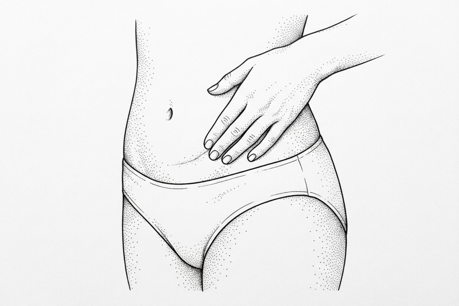
How a C-section incision heals
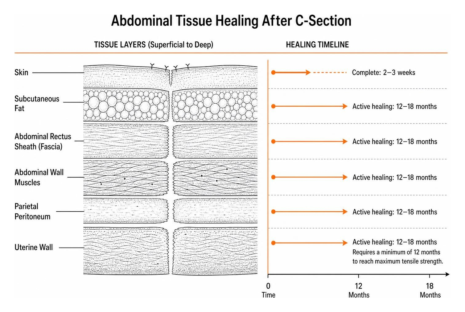
A surgical incision that penetrates the full depth of the abdominal wall cannot leave behind completely unblemished tissue. When evaluating whether surgical marks fade or disappear spontaneously, the biological reality is that scar tissue represents a permanent structural replacement for normal skin. While a visible mark will almost always remain, its prominence, color, and texture transform over an extended timeline.
The body heals a cesarean delivery across several anatomical layers: the outer skin, subcutaneous fat, the abdominal rectus sheath (fascia), the abdominal wall muscles, the parietal peritoneum, and the uterine wall. Fertilia reports that surface skin closure generally finishes within two to three weeks, yet the deeper fascial and uterine layers continue active repair and remodeling for 12 to 18 months. The uterine muscle layer requires a minimum of twelve full months to achieve its maximum tensile strength.
Determining when to start scar treatment after surgery depends on internal versus external tissue progress. The entire wound maturation cycle often extends up to two full years post-delivery, meaning early post-operative firmness, pinkness, or raised borders do not represent the final appearance.
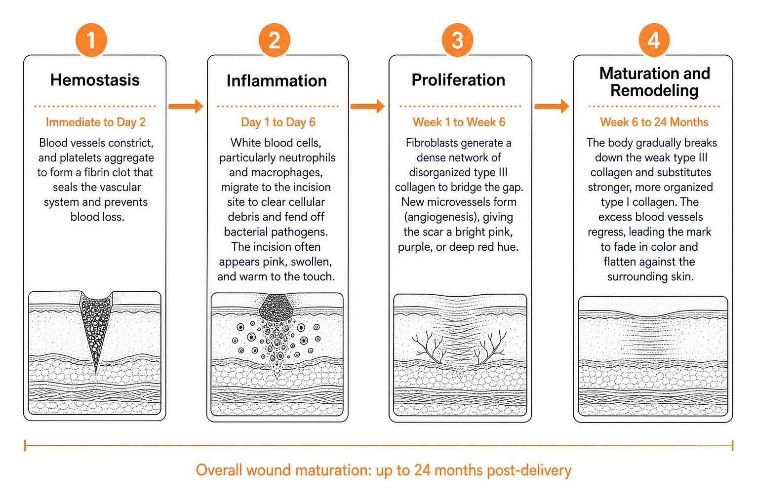
The stages of C-section healing
The recovery of an abdominal incision follows four overlapping biological stages:
- Hemostasis (Immediate to Day 2): Blood vessels constrict, and platelets aggregate to form a fibrin clot that seals the vascular system and prevents blood loss.
- Inflammation (Day 1 to Day 6): White blood cells, particularly neutrophils and macrophages, migrate to the incision site to clear cellular debris and fend off bacterial pathogens. The incision often appears pink, swollen, and warm to the touch.
- Proliferation (Week 1 to Week 6): Fibroblasts generate a dense network of disorganized type III collagen to bridge the gap. New microvessels form (angiogenesis), giving the scar a bright pink, purple, or deep red hue.
- Maturation and Remodeling (Week 6 to 24 Months): The body gradually breaks down the weak type III collagen and substitutes stronger, more organized type I collagen. The excess blood vessels regress, leading the mark to fade in color and flatten against the surrounding skin.
What affects how fast a scar matures
The rate of scar fading varies based on surgical technique, individual genetics, and physical stress placed on the incision. Incision orientation plays a substantial role. A horizontal low-transverse incision (the standard "bikini cut") runs parallel to natural skin tension lines (Langer's lines), minimizing mechanical strain during movement and promoting smoother, thinner healing lines.
In contrast, a midline vertical incision runs across these tension lines, subjecting the healing edges to continuous lateral pull from the abdominal wall. In a clinical study reported by Rub-Maps evaluating abdominal scar progression, vertical midline incisions showed an odds ratio of 20.53 for developing hard, raised hypertrophic scarring compared to low transverse incisions.
Other biological and lifestyle factors include:
- Melanin Levels: Individuals with deeper skin tones frequently experience prolonged post-inflammatory hyperpigmentation or hypopigmentation, leaving brown, dark purple, or white marks that take longer to blend.
- Tissue Tension and Movement: Early strenuous lifting or persistent coughing strains the deep fascial layer, prompting fibroblasts to synthesize extra collagen.
- Systemic Health: Conditions like diabetes, metabolic stress, and poor microcirculation slow the delivery of oxygen and nutrients needed for collagen remodeling.
- Wound Complications: Postoperative infections or small areas of wound separation extend the inflammatory phase, which stimulates excess scar tissue formation.
Types of C-section scars
| Scar Category | Visual Appearance | Texture & Elevation | Typical Timeline & Behavior |
|---|---|---|---|
| Normal / Mature | Pale, thin line matching or slightly lighter than skin tone | Flat, soft, flexible | Matures over 12–24 months; rarely causes functional discomfort |
| Hypertrophic | Red, pink, or purplish; strictly stays within incision margins | Raised, rigid, thick | According to Rub-Maps, present in 64.3% of patients at 3 months and drops to 35.3% at 1 year |
| Keloid | Deep red, dark brown, or hyperpigmented; expands beyond the original cut | Firm, bulbous, nodular | Behaves like a benign growth; rarely regresses without intervention |
| Tethered (Shelf) | Indented line with an overhanging fold of skin/fat | Dense beneath the skin; anchored to muscle | Results from adhesions binding dermis to deep fascia; improves with manual mobilization |
A broader scar types overview details morphological distinctions across abdominal incisions.
Hypertrophic scars vs. keloids
Hypertrophic scars and keloids are both driven by an overproduction of collagen, but their clinical behaviors diverge significantly. Hypertrophic scars remain confined within the boundaries of the original surgical incision. They often appear red, firm, and elevated during the first three to six months before gradually softening.
A keloid, however, extends outward into previously undamaged tissue, forming irregular, rubbery mounds that can cause intense itching, burning, and localized pain. Keloids do not regress naturally. When elevated scars resist topical management, clinicians frequently use steroid injections for surgical and c-section scars to inhibit fibroblast hyperactivity, break down collagen bonds, and reduce local swelling.
Why the scar can tether and form a shelf
Many patients observe a physical fold or "shelf" of soft tissue hanging over the incision line. While often mistaken for abdominal adipose tissue, this feature frequently stems from deep tissue tethering.
During surgery, multiple anatomical layers are incised and sutured. As these layers heal, dense fibrous adhesions can bind the underside of the skin directly to the underlying muscle fascia. This anchors the scar inward while the unattached subcutaneous fat above it rests forward.
Flexa Fit describes localized swelling from interrupted lymphatic channels as another contributor to this contour discrepancy during early postpartum recovery. Over the course of the first year, manual desensitization and guided soft-tissue release can improve tissue glide and soften the appearance of the shelf.
Non-Invasive Management and Home-Based Scar Mobilization
Supporting optimal wound maturation involves reducing tension, maintaining skin barrier hydration, and encouraging healthy collagen alignment.
Scar massage and myofascial release
Targeted massage prevents deep scar tissue from adhering to the underlying abdominal muscles. Gentle desensitization around the surrounding skin can begin around six weeks postpartum once the incision is fully closed, dry, and cleared by a physician.
- Weeks 6 to 8 (Desensitization): Using clean hands and a few drops of unscented oil, make light circular motions on the intact skin 1 to 2 inches above and below the incision line to re-educate cut sensory nerves.
- Months 2 to 3 (Direct Mobilization): Place the pads of two fingers directly over the healed scar line. Apply gentle downward, upward, and circular pressure to shift the skin over the deep fascia without causing pain.
- Months 3 to 6 (Cross-Fiber Friction & Rolling): Apply gentle cross-fiber friction by moving fingers perpendicular to the incision line, or gently lift the skin between the thumb and forefinger to release deeper fascial restrictions.
Topical hydration outlined in the postpartum scar reduction cream guide provides non-irritating barrier support during mobilization.
Medical-Grade Silicone Sheeting and Gels
Topical silicone remains a standard at-home strategy for managing raised or pigmented incisions. Silicone sheets and gels create a semi-occlusive barrier that mimics the stratum corneum, locking in moisture and normalizing capillary activity. This hydration signals fibroblasts to moderate collagen production, helping to soften and flatten the scar line.
Consistent wear for 12 to 24 hours daily over a span of two to four months produces optimal outcomes, following standard protocols in the non-invasive C-section scars guide.
Treatments for stubborn C-section scars
When textural irregularities, hyperpigmentation, or tissue tethering persist beyond the initial year of healing, advanced clinical interventions provide aesthetic and functional improvements.
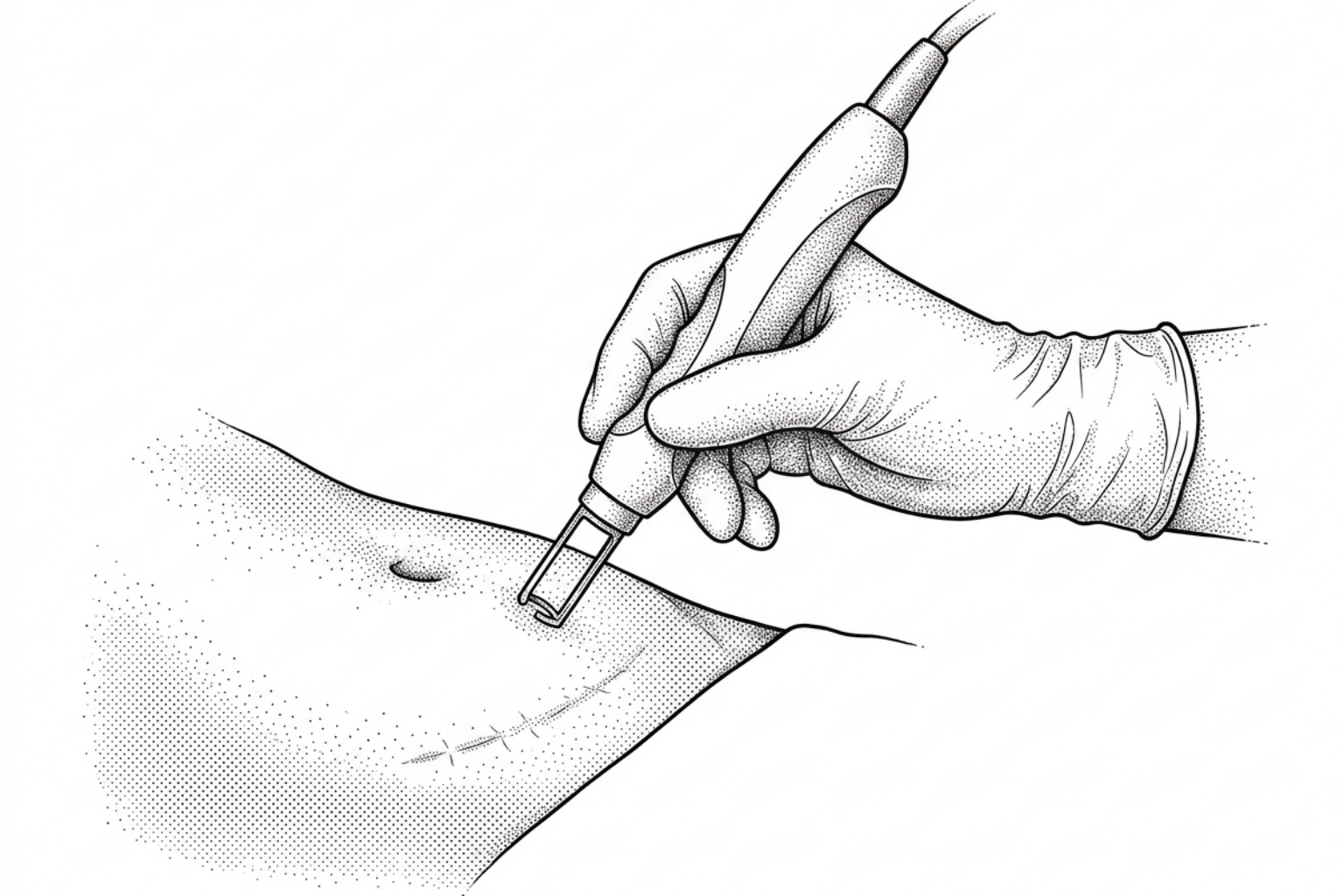
Lasers and subcision
Dermatologists and plastic surgeons utilize various energy modalities to remodel established scar tissue:
- Vascular Lasers (Pulsed Dye Laser): Target hemoglobin in superficial blood vessels to diminish lingering erythema or purple discoloration in active scars.
- Fractional CO2 Laser Resurfacing: Delivers microscopic columns of thermal energy into the dermis to break up disorganized collagen fibers and stimulate organized skin regeneration. Clinicians apply C-section scar laser resurfacing alongside broader protocols for laser treatment for surgical scars.
- Subcision: For tethered incisions or deep scar shelves, a clinician inserts a specialized needle beneath the skin under local anesthesia to physically sever the fibrous bands anchoring the dermal layer to the deep muscle fascia.
Surgical scar revision
For wide, asymmetric, or severely restricted marks that do not respond to non-invasive therapies, surgical revision remains an option. A surgeon excises the old fibrous scar tissue and re-closes the wound under minimal tension using layered dermal suturing techniques. Detailed surgical pathways are discussed in the C-section scar revision guide and the general surgical scar revision guide.
Normal healing vs. problems to watch for
Recognizing typical healing sensations versus clinical complications ensures appropriate care throughout the postpartum period.
What normal scar sensations feel like
- Mild Itching: Often caused by skin stretching, tissue repair, and local hair regrowth along the pubic line.
- Surface Numbness: Cutaneous sensory nerve branches severed during surgery regenerate at approximately 1 millimeter per day, so localized numbness may persist for 6 to 12 months.
- Transient Aching: Occasional twinges or pulling sensations during physical activity or core engagement as the fascia adjusts to mechanical load.
Signs you should call your doctor
- Spreading redness, localized heat, or swelling around the wound edges.
- Yellow, green, or foul-smelling drainage from the incision site.
- Persistent maternal fever above 100.4°F (38°C).
- Any separation or opening (dehiscence) along the incision margins.
- A painful, firm nodule that swells or bleeds in rhythm with the menstrual cycle, which may indicate a scar endometrioma (endometrial tissue implanted into the abdominal wall).
- Heavy vaginal bleeding, chronic pelvic pain, or intermenstrual spotting, which can signal an isthmocele (a cesarean scar defect within the uterine wall). According to data from the Cleveland Clinic, an isthmocele occurs in an estimated 20% to 70% of women who undergo at least one cesarean delivery.
Common questions about C-section scars
Will C-section scars fade without treatment?
While a surgical mark does not vanish completely, most low-transverse cesarean incisions fade into a thin, pale, flat line over 12 to 24 months without advanced medical intervention. In many instances, the mature scar sits below the underwear line and is naturally obscured by pubic hair.
Why pain and numbness can linger
Prolonged numbness or intermittent sharp twinges occur because tiny peripheral nerve branches in the skin were cut during the procedure. As these sensory nerves slowly regenerate, they can cause sensations of tingling, burning, or dull numbness. Persistent, deep aching months later may also stem from scar tissue adhering to the abdominal wall muscles.
Why a firm lump or overhang forms
A hard ridge under the scar during the first two to three months usually represents a temporary buildup of early, disorganized type III collagen, which softens as remodeling advances. A visible shelf or overhang is often caused by fascial tethering (where the scar is anchored firmly to deep muscle) combined with localized fluid accumulation and natural subcutaneous tissue distribution.
Conclusion
Understanding how abdominal tissue repairs itself helps set realistic expectations for the postpartum recovery journey. A cesarean scar transforms through distinct biological stages over a multi-year timeline. While the marks left by major surgery never disappear entirely, consistent scar mobilization, sun protection, and medical-grade barrier therapies can dramatically improve comfort and cosmetic appearance.
For individualized treatment comparisons and step-by-step care pathways, visit the complete C-section scar hub.
This content is for informational purposes only and does not constitute medical advice. Consult a qualified healthcare professional for diagnosis and treatment.
Works Cited
- Cleveland Clinic. "Isthmocele (Cesarean Scar Defect): Symptoms, Causes, and Treatment." Cleveland Clinic Health Library, 2024.
- Fertilia. "C-Section Scar: Healing Timeline, Massage & Signs to Watch." Fertilia Clinical Insights, 2026.
- Flexa Fit. "C Section Scar Recovery: Healing, Massage and What Is Normal." Flexa Fit Health & Movement, 2026.
- Rub-Maps. "Why 1 In 3 C-Section Scars Turn Hard and Raised – and What the Dressing Trials Reveal." Rub-Maps Clinical Analysis, 2026.