The Art of Scar Camouflage and Color Correction
Dark scars can be stubborn, but the right approach depends on what is causing the discoloration. Explore evidence-based methods for dark scar removal and color correction.

What Clinical Evidence Says About Dark Scar Removal Methods
Dark scar removal methods range from topical agents to laser-based procedures, and the right approach depends on scar type, skin tone, and the underlying biology driving the discoloration.
Quick reference — evidence-based dark scar removal methods:
| Method | Best For | Evidence Level |
|---|---|---|
| Laser therapy (PDL, Nd:YAG, fractional CO2) | Raised, pigmented, or textured scars | Strong clinical evidence |
| Intralesional corticosteroid injections | Keloid and hypertrophic scars | Strong; 50–100% response rate |
| Topical hydroquinone / retinoids | Post-inflammatory hyperpigmentation | Moderate to strong |
| Silicone gel sheets | Prevention and flattening of raised scars | Strong; supported by clinical guidelines |
| Cryosurgery | Keloid and hypertrophic scars | Moderate; 20–75% volume reduction |
| Chemical peels | Surface pigmentation, atrophic scars | Moderate |
| Microneedling | Atrophic scars, collagen remodeling | Moderate |
| Sun protection (SPF 30+) | Preventing further darkening | Strong; essential adjunct |
Scars are a normal outcome of the skin's repair process. When the dermis — the deeper layer of skin — sustains damage, the body lays down collagen fibers to close the wound. That process is efficient, but imperfect. The result is tissue that looks and behaves differently from the surrounding skin.
For many people, the concern isn't just the scar's texture. It's the color.
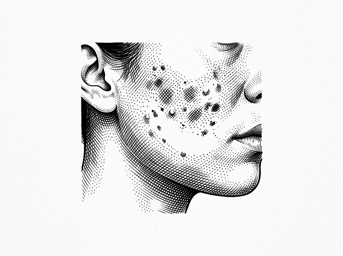
Dark or hyperpigmented scars are especially common in people with medium to deep skin tones — those with African, Asian, or Hispanic heritage in particular. This is partly because melanocytes, the cells that produce skin pigment, are more reactive following inflammation. The result is a condition called post-inflammatory hyperpigmentation (PIH) — a darkening of the skin that can persist long after the original wound has closed.
The challenge is real. Keloids, for example, develop in up to 6–16% of people with African, Asian, or Hispanic heritage. Hypertrophic scars following burns occur in up to 78% of cases. And unlike lighter skin tones, where scars often appear pink or red, darker skin tones tend to show scars as brown or near-black spots — sometimes more visually prominent than the structural scar itself.
Treatment is possible. But it requires matching the right method to the right scar type — and understanding how different interventions interact with melanin-rich skin before starting.
Terms related to dark scar removal methods:
Pathophysiology of Hyperpigmentation in Dermal Scarring
The biological mechanism behind a dark scar begins with the inflammatory phase of wound healing. When the skin is breached, the immune system releases cytokines and inflammatory mediators. In individuals with higher baseline melanin—specifically those categorized as Fitzpatrick skin types IV through VI—these signals do more than just recruit repair cells; they also stimulate melanocytes.
Melanocytes are the pigment-producing cells located at the basal layer of the epidermis. During the repair process, an overabundance of melanin can be transferred to neighboring keratinocytes or even "leak" into the dermis, where it becomes trapped. This process, known as melanogenesis, is the engine behind post-inflammatory hyperpigmentation (PIH).
Research indicates that the depth of the pigment dictates the difficulty of treatment. Epidermal PIH (surface-level) often responds well to topical interventions, while dermal PIH (deep-level) may require energy-based devices to reach the trapped pigment. Furthermore, in scarred tissue, the irregular arrangement of collagen fibers can alter how light reflects off the skin, making the dark pigment appear even more pronounced or uneven.
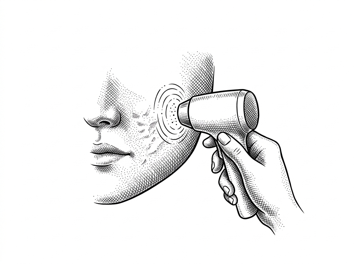
Clinical Efficacy of Laser and Light-Based Dark Scar Removal Methods
Laser technology has transformed the landscape of dark scar removal methods by utilizing the principle of selective photothermolysis. This involves using specific wavelengths of light to target a "chromophore"—in this case, melanin or hemoglobin—without damaging the surrounding healthy tissue.
For pigmented scars, lasers work by breaking down the excess melanin into smaller particles that the body's lymphatic system can then clear away. This is detailed extensively in the Laser Treatment for Scars Complete Guide. However, clinicians must exercise caution with darker skin tones, as the laser may inadvertently target the skin's natural pigment, leading to hypopigmentation (white spots) or worsening hyperpigmentation.
A standard approach for structural scars that also exhibit dark pigment is the use of the CO2 Laser Scar Removal technique. This ablative method removes thin layers of skin while heating the deeper dermis to stimulate new, organized collagen growth, effectively "resetting" the skin surface.
Targeted Wavelengths for Dark Scar Removal Methods
Different colors in a scar require different wavelengths:
- 595 nm (Pulsed Dye Laser): Primarily used for the vascular component of scars. While often associated with redness, the Red Scar Fading Laser can also help reduce the inflammatory signals that lead to subsequent darkening.
- 1064 nm (Nd:YAG): This longer wavelength penetrates deeper and is generally safer for darker skin tones because it is less absorbed by surface melanin, reducing the risk of burns.
- Q-switched and Picosecond Lasers: These deliver energy in incredibly short bursts, creating a mechanical effect that shatters pigment particles. Studies show picosecond lasers can achieve 32% to 45% scar volume reduction in atrophic scars while addressing dyschromia.
Fractional Photothermolysis for Pigment Correction
Fractional technology creates thousands of microscopic treatment zones (MTZs) while leaving the surrounding skin intact. This "bridge" of healthy skin allows for much faster healing and a lower risk profile. For surgical marks, viewing CO2 Laser for Surgical Scars Before and After results often reveals significant improvement in both the "ropiness" of the scar and its dark coloration. This method essentially forces the skin to turn over its pigment-heavy cells and replace them with normally pigmented tissue.
Pharmacological and Topical Interventions for Pigmented Scars
Topical treatments are often the first line of defense, especially for flat, dark scars. The most effective agents are tyrosinase inhibitors, which block the enzyme responsible for melanin production.
Hydroquinone remains the gold standard for lightening dark spots, often used in concentrations of 2% (over-the-counter) to 4% (prescription). However, it is generally recommended for short-term use (3-6 months) to avoid a rare side effect called ochronosis. For older marks, a specialized old scar lightening serum often combines hydroquinone with other brighteners like kojic acid or vitamin C.
Retinoids (such as tretinoin) are also vital. They speed up cell turnover, pushing pigmented cells to the surface to be shed, and improve the penetration of other lightening agents. This is a core component of many quick scar fading treatments.
| Ingredient | Mechanism | Efficacy for Dark Scars |
|---|---|---|
| Hydroquinone | Tyrosinase inhibitor | High (Gold Standard) |
| Azelaic Acid | Selective for hyperactive melanocytes | Moderate to High |
| Vitamin C | Antioxidant; inhibits melanin synthesis | Moderate (Best for prevention) |
| Glycolic Acid | Exfoliation; increases cell turnover | Moderate (Adjunctive) |
| Niacinamide | Blocks pigment transfer to cells | Moderate |
Managing Keloids and Hypertrophic Scars in Pigmented Skin
Raised scars present a dual challenge: they are structurally abnormal and frequently hyperpigmented. Keloids are particularly aggressive, as they grow beyond the boundaries of the original wound and continue to produce excess collagen for years.
The management of keloid disease in ethnic skin requires a structured approach. Intralesional corticosteroid injections (such as triamcinolone acetonide) are the most common treatment. These injections work by suppressing inflammation and inhibiting fibroblast proliferation—the cells responsible for the excess collagen. Clinical data shows these injections can reduce scar size by 50% or more, and in some cases, make the scar appear almost flat.
For stubborn cases, combination therapy is often necessary. This might include:
- Corticosteroids + 5-Fluorouracil (5-FU): An anti-metabolite that further slows cell growth.
- Cryosurgery: Freezing the scar tissue. Intralesional cryosurgery, where the needle freezes the scar from the inside out, has shown a 20% to 75% reduction in volume and is less likely to cause the "white spots" associated with surface freezing.
- Laser Revision: Following the flattening of a scar, Surgical Scar Laser Treatment can be used to address the remaining dark pigment.
Evidence-Based Strategies for Preventing Post-Surgical Dyschromia
Prevention is significantly more effective than removal. The goal post-surgery or post-injury is to minimize inflammation and tension, both of which trigger excess collagen and pigment.
Silicone gel and silicone gel sheets have been used since 1982 and remain a cornerstone of scar management. They work through occlusion and hydration, which signals the skin to "slow down" collagen production. Evidence suggests that applying silicone sheets daily as soon as the wound has closed can significantly reduce the incidence of hypertrophic scarring.
Early intervention with light-based devices is also gaining traction. Using Laser Treatment for Surgical Scars within the first few weeks of healing can prevent the formation of the dense, dark tissue. For those dealing with the aftermath of acne, the goal is to reduce acne scars fast by treating active inflammation immediately to prevent the PIH cycle from ever starting.
The Role of Photoprotection in Dark Scar Removal Methods
Sunlight is the enemy of scar healing. Ultraviolet (UV) radiation directly stimulates melanocytes to produce more pigment. A healing scar that is exposed to the sun is much more likely to darken permanently.
Medical consensus dictates the use of a broad-spectrum SPF 30+ sunscreen. Mineral blockers containing titanium dioxide or zinc oxide are often preferred for scarred skin because they sit on top of the skin and reflect UV rays without causing the potential irritation sometimes associated with chemical filters. Protection should be maintained for at least 12 to 18 months—the time it typically takes for a scar to fully mature.
Adjunctive Natural Compounds and Evidence Limitations
While the internet is full of "miracle" cures, clinical evidence for many natural remedies is thin.
- Aloe Vera: A 2019 review of 23 trials suggests aloe vera can aid wound healing and hydration, which may indirectly improve scar appearance. It is a safe, hydrating adjunct, as noted in research on aloe vera scar reduction.
- Vitamin E: Despite its popularity, a 2016 review concluded there is insufficient evidence that topical vitamin E improves scars. In some cases, it can even cause contact dermatitis (an itchy rash).
- Honey: While medical-grade honey has antibacterial properties that help wounds heal, a 2016 study found it did not significantly improve the cosmetic appearance of existing scars.
Frequently Asked Questions about Pigmented Scars
How long does it typically take for dark scars to fade with clinical treatment?
Patience is essential. Most scars take 12 to 18 months to mature naturally. With clinical intervention, such as lasers or chemical peels, initial pigment fading may be seen in 3 to 6 months, but structural remodeling and full color correction often require multiple sessions spaced 4 to 8 weeks apart.
Are natural remedies like honey or onion extract effective for color correction?
Onion extract has shown some promise in clinical studies for improving the texture and itchiness of keloids, but its effect on deep pigmentation is limited. Honey is excellent for preventing infection in open wounds but has little evidence for removing established dark pigment.
When is a professional dermatological consultation necessary for scar management?
You should see a board-certified dermatologist if the scar is growing, painful, or restricts movement. Furthermore, because some skin cancers can mimic the appearance of a scar, any new or changing "scar" that appeared without a clear injury should be evaluated immediately.
Conclusion
Successfully navigating dark scar removal methods requires a blend of biological understanding and clinical precision. Whether through the use of advanced lasers, targeted tyrosinase inhibitors, or mechanical therapies like silicone sheeting, the goal is to guide the skin back to its natural state. While no scar can be "erased" 100%, modern dermatology offers powerful tools to camouflage and correct even the most stubborn pigmentation. For the latest in skin regeneration research, subscribe for more research-backed skin health updates.
Works Cited
- Davis EC, Callender VD. Postinflammatory hyperpigmentation: a review of the epidemiology, clinical features, and treatment options in skin of color. J Clin Aesthet Dermatol. 2010;3(7):20-31.
- Ud-Din S, Bayat A. Strategic management of keloid disease in ethnic skin: a structured approach supported by the emerging literature. Br J Dermatol. 2013;169:71–81.
- Gold MH, et al. Updated international clinical recommendations on scar management: part 2--algorithms for scar prevention and treatment. Dermatol Surg. 2014;40(8):825-31.
- O'Connor K, et al. Laser and light-based treatments for scars: a review. J Clin Aesthet Dermatol. 2020;13(11):34-41.
- Andrews JP, et al. Keloids: the paradigm of skin fibrosis—pathomechanisms and treatment. Matrix Biol. 2016;51:37–46.
This content is for informational purposes only and does not constitute medical advice. Consult a qualified healthcare professional for diagnosis and treatment.